A 19-year-old male patient presented with a longstanding complaint of decreased vision. He had been diagnosed with retinitis pigmentosa at the age of 10. The patient also reported congenital hearing impairment. Family history was remarkable for retinitis pigmentosa in his mother and hearing impairment in his maternal grandmother.
On ophthalmological examination, the best-corrected visual acuity was 0.9 in the right eye and 0.4 in the left eye. Intraocular pressure was measured as 17 mmHg in the right eye and 22 mmHg in the left eye. Slit-lamp examination revealed posterior subcapsular cataracts in both eyes. Fundus examination revealed bilaterally pale optic discs, attenuated retinal vessels, and bone spicule–like pigmentary changes in the midperipheral retina. These findings were consistent with Usher syndrome.


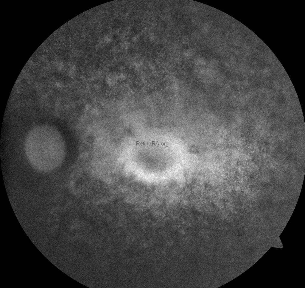
On color fundus photography, retinitis pigmentosa is typically characterized by the presence of bone spicule (showed by arrow), attenuation of the retinal arterioles, and waxy pallor of the optic disc. In advanced stages like our case macular involvement is observed, including cystoid macular edema.


On fundus autofluorescence imaging, the patient exhibited a parafoveal hyperautofluorescent ring, delineating the border between relatively preserved and degenerated photoreceptor regions. Beyond this ring, patchy hypoautofluorescent areas were observed in the midperipheral retina, consistent with widespread retinal pigment epithelium and photoreceptor loss. These findings are characteristic of retinitis pigmentosa.


Optical coherence tomography (OCT) demonstrated cystoid macular edema characterized by intraretinal cystic spaces in the foveal region. An epiretinal membrane was also observed, contributing to foveal contour irregularity. The outer retinal layers, particularly the ellipsoid zone, appeared disrupted and thinned, consistent with advanced photoreceptor degeneration. These findings are in line with retinal structural changes observed in Usher syndrome.
Usher syndrome is a rare autosomal recessive disorder characterized by the combination of sensorineural hearing loss and progressive retinal degeneration, most commonly retinitis pigmentosa. It represents the most frequent cause of combined deafness and blindness. The clinical spectrum is heterogeneous and has been classified into three major types based on the severity and onset of auditory and vestibular dysfunction. Type I is associated with congenital profound deafness, vestibular areflexia, and early-onset retinitis pigmentosa; Type II presents with moderate-to-severe congenital hearing loss, intact vestibular function, and later-onset retinitis pigmentosa; and Type III is characterized by progressive hearing loss, variable vestibular dysfunction, and variable age of onset of retinitis pigmentosa.
The underlying pathology of Usher syndrome involves mutations in genes critical for the development and maintenance of photoreceptors and cochlear hair cells. Clinically, patients initially present with night blindness and peripheral visual field constriction due to retinitis pigmentosa, alongside varying degrees of hearing impairment. Over time, these changes lead to tunnel vision and eventual central vision loss. Although there is currently no curative treatment, genetic testing, low-vision rehabilitation, hearing aids or cochlear implants, and potential future gene therapies represent important aspects of patient management.
Cystoid macular edema in Usher syndrome and retinitis pigmentosa is typically managed with topical or oral carbonic anhydrase inhibitors, which represent the first-line therapy. In refractory cases, corticosteroids or, less effectively, anti-VEGF agents may be considered. Surgical options such as vitrectomy with epiretinal membrane peeling can be beneficial in patients with significant tractional components. However, the overall prognosis remains guarded, as CME often recurs and is influenced by the underlying photoreceptor degeneration.
Credit: M. Giray Ersoz, MD, FEBO
Biruni University School of Medicine, Department of Ophthalmology, Istanbul, Turkey
Instagram accounts: @retina.review and @retina.dr.girayersoz
and Sepideh Lotfi, MD
Biruni University School of Medicine, Department of Ophthalmology, Istanbul, Turkey
Instagram accounts: @sepidls