Torpedo maculopathy was detected in the routine eye examination of an 18-year-old female patient. Visual acuity in both eyes was 20/20.
Torpedo maculopathy, also known as solitary hypopigmented nevus of the retinal pigment epithelium (RPE), paramacular albinotic spot syndrome, congenital hypomelanotic freckle, or atypical macular coloboma was first described by Roseman and Gass in 1992 as a rare congenital anomaly of the RPE that produces a disruption of outer retinal layers. So far, short series and scarcely any data about prevalence, demographics or incidence have been reported. Pathogenesis remains unknown and the typical lesion is a single hypopigmented area in the macula, asymptomatic, temporal to fovea and with a characteristic torpedo-shape. The lesion and visual acuity are usually stable. However, due to the possibility of CNV development and rare progression, follow-up at certain intervals is recommended.
Credit: M. Giray Ersoz, MD, FEBO
Biruni University School of Medicine, Department of Ophthalmology, Istanbul, Turkey
Instagram accounts: @retina.review and @retina.dr.girayersoz
Color Photography of Torpedo Maculopathy 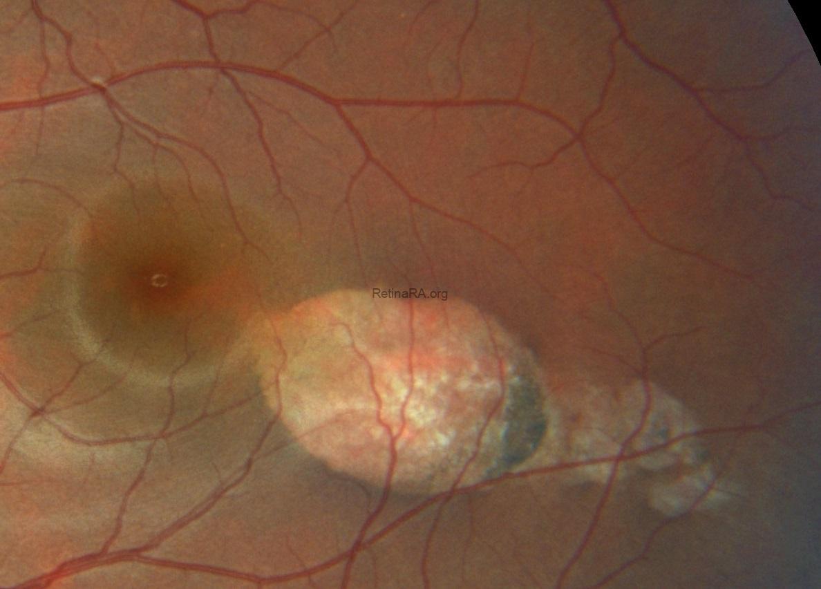
Autofluorescence Imaging of Torpedo Maculopathy 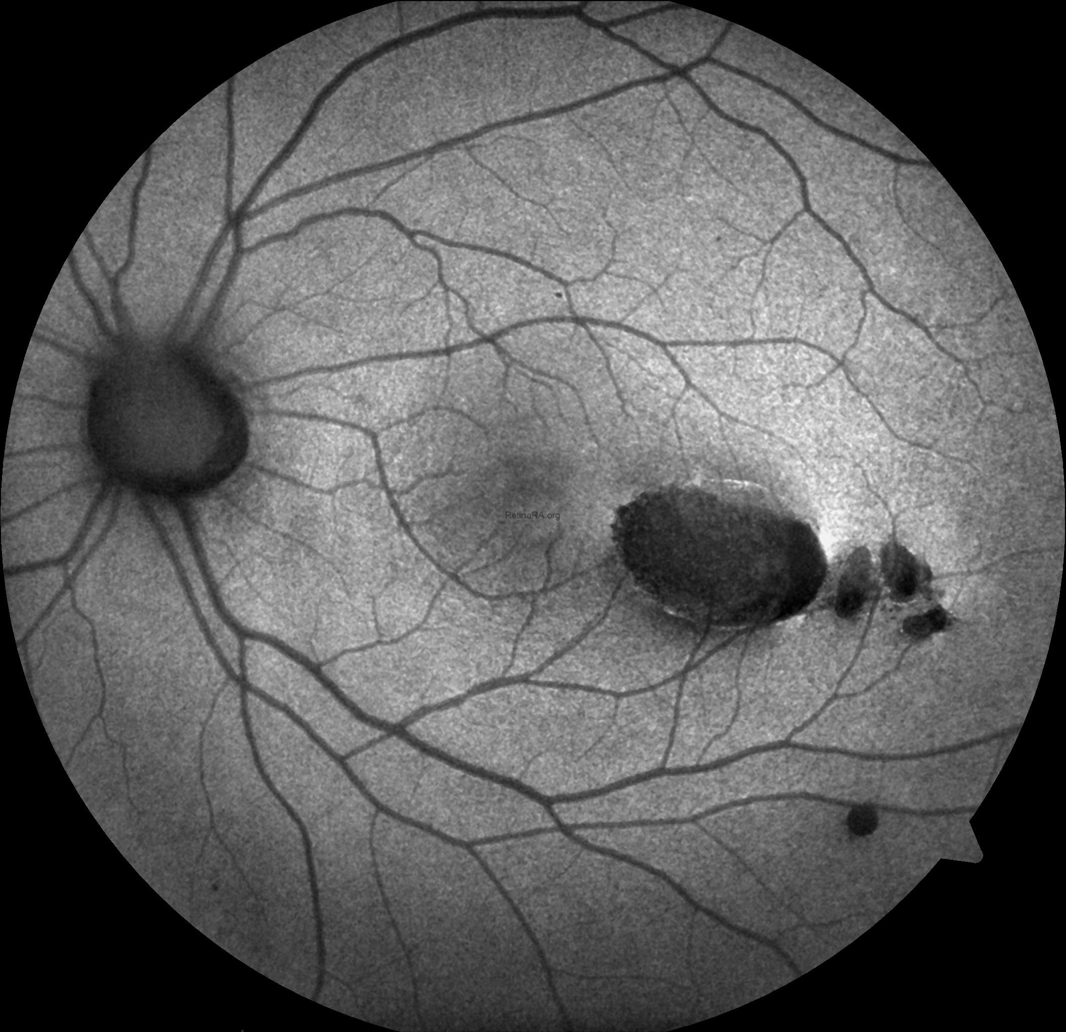
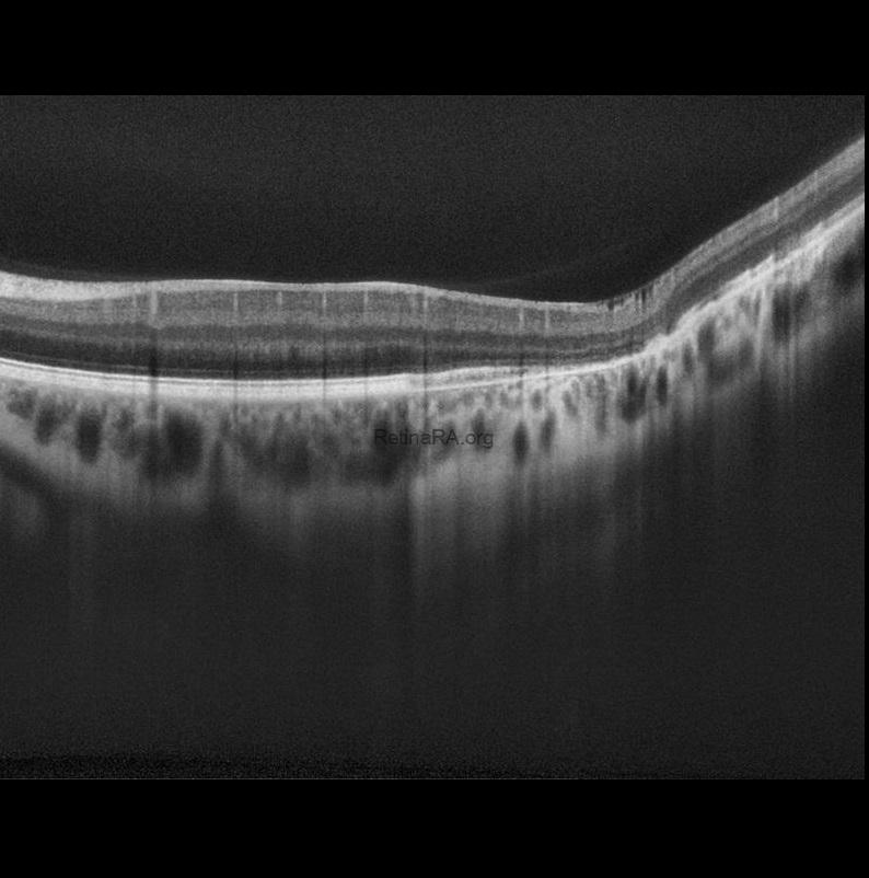
OCT of Torpedo Maculopathy