
A 22-year-old heathy woman applied to the outpatient ophthalmology clinic for a routine eye examination. The BCVAs were 20/20 for both eyes and IOPs were within normal limits. Anterior segment examination was also unremerkable.
Fundus examination showed the sharply demarcated bands of tightly packed groups of glistening white dots like snowflakes that give the superior temporal periphery of the retina a white frost-like appearance.
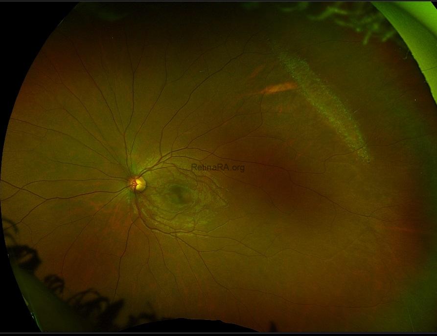
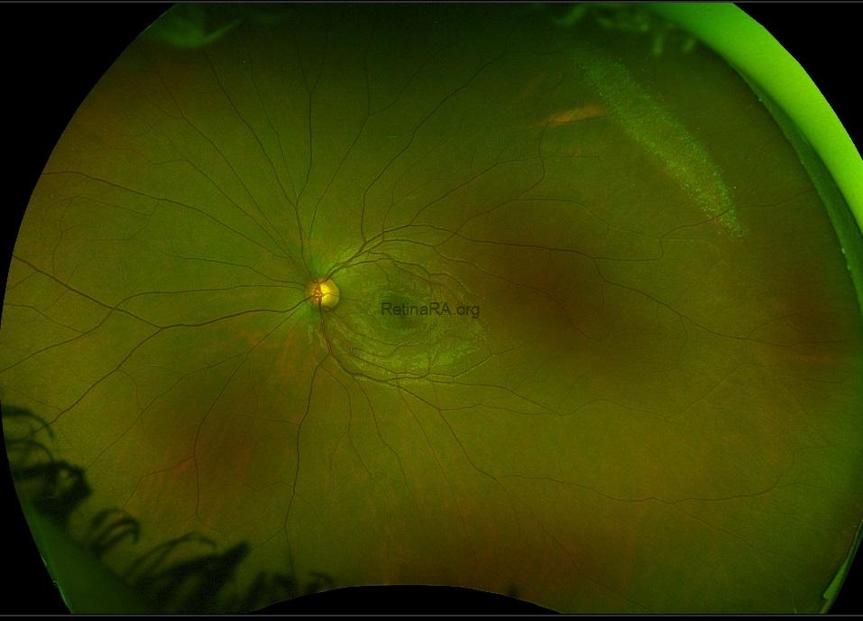
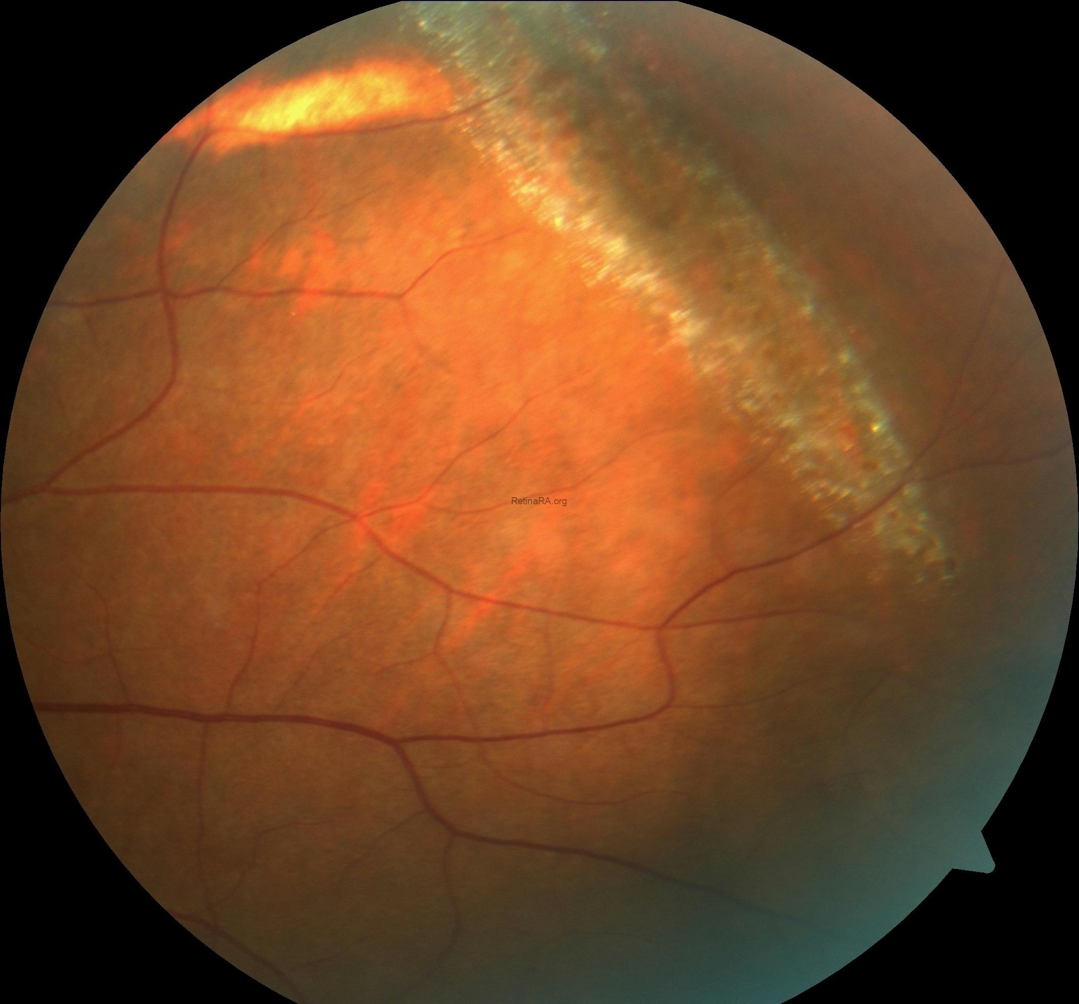
Snail-track degeneration of retina is a glistening white frost like area, elongated oval or spindle shaped and has multiple white dots within it. Snail-track degenration is found in approximately 10% of the general population and more common in myopic eyes (up to 40%). It usually seen in the superotemporal and superonasal quadrants and the majority seen at least 2 disk diameter anterior to the equator. Snail track degeneration can resemble lattice degeneration and may sometimes be noted as early lattice. Marked vitreous traction is seldom present in it so that U-tears rarely occur, although round holes are relatively common. Prophylactic treatment is not necessary.
Credit: Kemal Tekin, M.D., from Ulucanlar Eye Training and Research Hospital
Instagram accounts: @retina.academy and @dr.kemaltekin