
A retinal arterial macroaneurysm refers to a localized dilation or ballooning of a retinal blood vessel, typically an arteriole, resulting in leakage or rupture of the vessel. It can lead to retinal hemorrhages, exudates, and edema, often affecting the macula. This condition is most commonly found in older adults with HT and can impair vision if not managed properly.
Color image, autofluorescence image, early stage of fluorescein angiography, and late stage of fluorescein angiography (respectively). 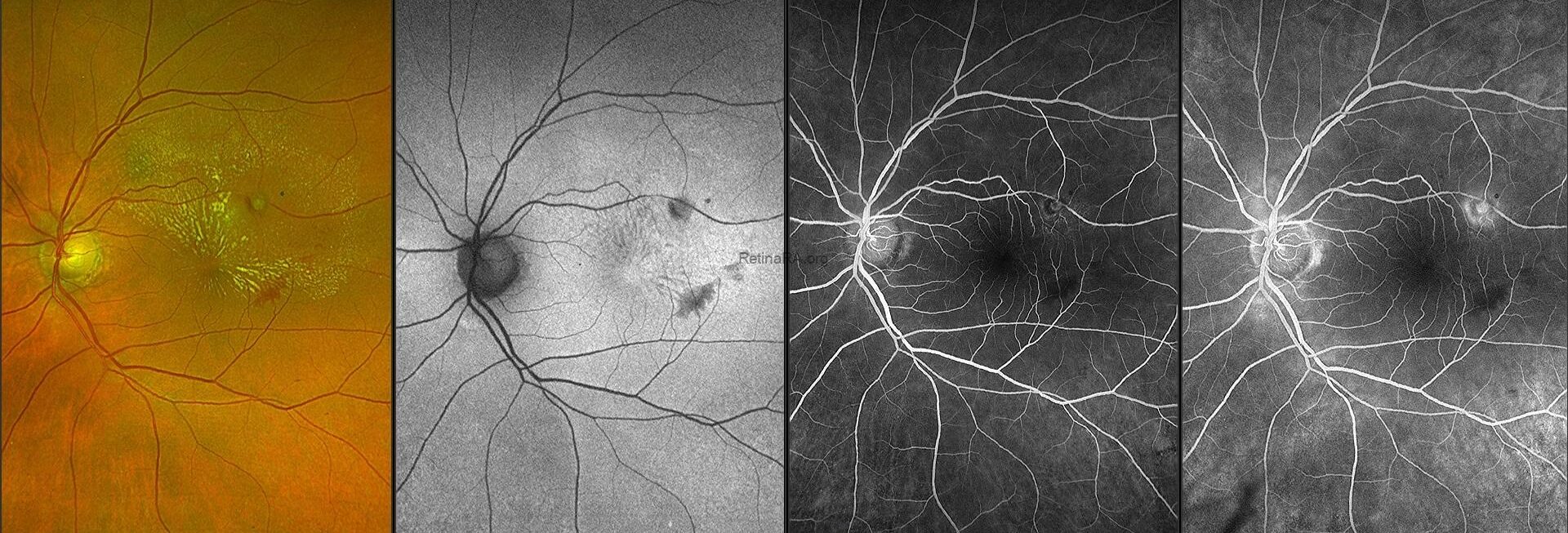
The retinal macroaneurysm typically presents as a round, well-circumscribed hyperfluorescent spot with progressive leakage seen in later phases of fluorescein angiography.
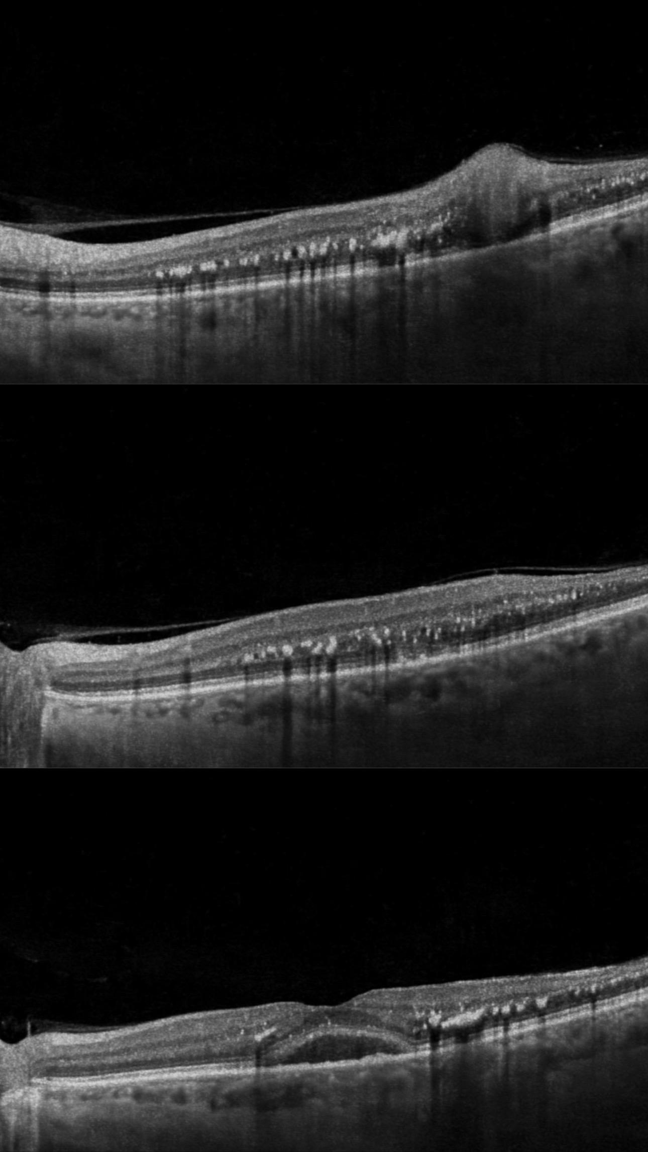
Optical Coherence Tomography (OCT) scans show :
-Elevated, round lesion at the inner retinal layers.
-Hyper-reflective dome-shaped appearance.
-Localized retinal thickening or
cystoid spaces.
-The hyperreflective lesion corresponding to exudates appears as bright, well-defined areas within the retina.
-Subretinal fluid in adjacent areas.
Credit: Dr. Benmoussa N Houda
Benmoussa Ophthalmology Clinic, Constantine, Algeria
Instagram account: @dr.benmoussa_houda