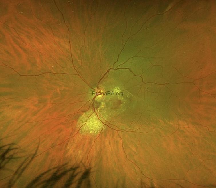
Polypoidal choroidal vasculopathy (PCV) was first described by Yannuzzi in 1982 as subretinal polypoidal vascular lesions associated with serous and hemorrhagic pigment epithelial detachments (PEDs). Recently, it has been suggested that its name be changed to aneurysmal type 1 macular neovascularization. It is a subtype of exudative age-related macular degeneration (AMD) and is also evaluated within the pachychoroid spectrum. The neovascular components of the PCV lesion include a polyp or polyps and a branching neovascular network.
Autofluorescence image (left side), reveals hypoautofluorescent area corresponding to hemorrhage, and this area is surrounded with gravitational slight hyperautofluorescence corresponding to chronic fluid accumulation. Fluorescein Angiography (FA) images depict early (middle) hyperfluorescence and late (right side) leakage from abnormal vessels (polyps and branching neovascular network).
 OCT reveals:
- A hemorrhagic PED with polyps (aneurysms): Hyporeflective lumen surrounded by a hyperreflective ring attached to the undersurface of RPE (top OCT image)
- Hyperreflective hemorrhage and hyporeflective fluid.
- Thick choroid with dilated Haller layer vessels (pachyvessels)
- Double-layer sign representing the branching neovascular network (bottom OCT image).

Credit: Dr. Benmoussa N Houda
Benmoussa Ophthalmology Clinic, Constantine, Algeria
Instagram account: @dr.benmoussa_houda
Caption editing: M. Giray Ersoz, MD, FEBO
Biruni University School of Medicine, Department of Ophthalmology, Istanbul, Turkey
Instagram accounts: @retina.review and @retina.dr.girayersoz