
This case was a 33-year-old male patient whose best corrected visual acuities were 20/25 for both eyes.
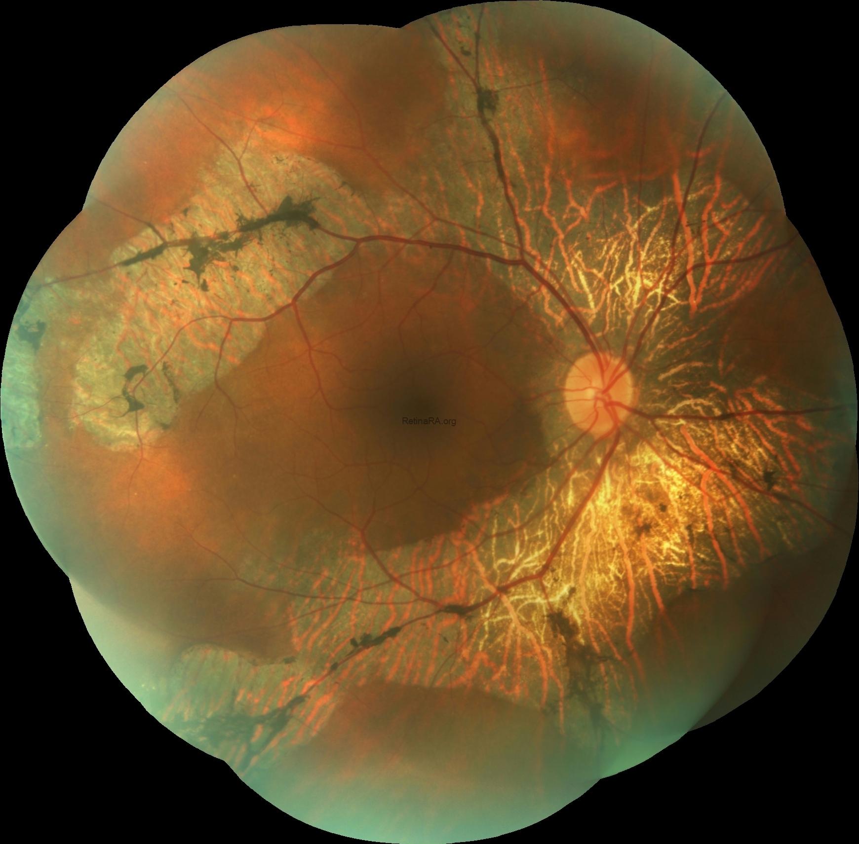
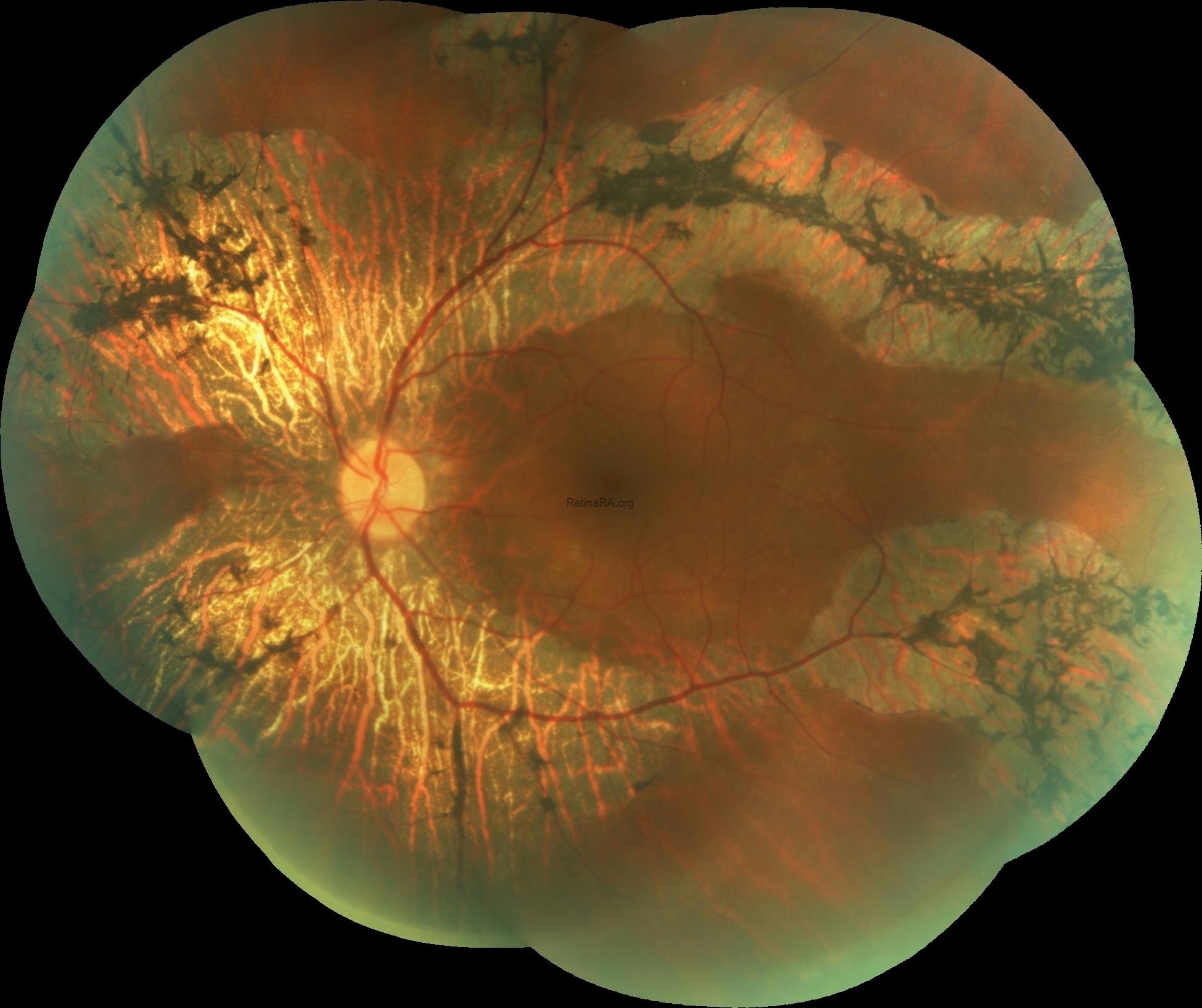
Fundus examination showed symmetrically-shaped bone-spicule pigmentation and retinochoroidal atrophy along the retinal veins in both eyes.
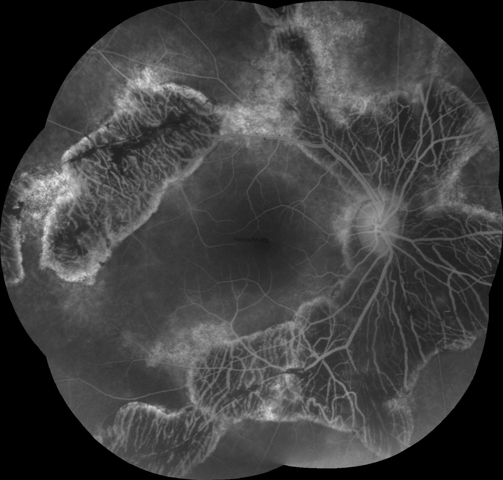
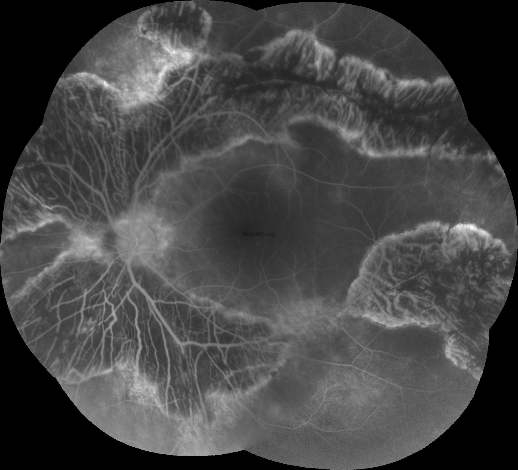
Fundus fluorescein angiography revealed the transmitted hyperfluorescence consistent with retinal pigment epithelium degeneration at the edges of atrophy in both eyes, with more extensive areas of choriocapillaris atrophy and blocked fluorescence in the pigment accumulation.
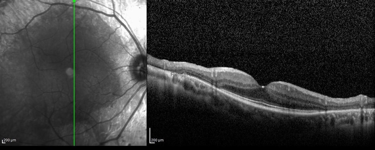
Vertical scans spectral-domain optical coherence tomography exhibited the atrophy of outer retinal layers and retina pigment epithelium at the paravenous areas with the normal retinal microstructure at macular areas in both eyes.

Pigmented paravenous retinochoroidal atrophy is characterized by perivenous aggregations of pigment clumps associated with peripapillary and radial zones of retinochoroidal atrophy which are distributed along the retinal veins. Patients are often asymptomatic and diagnosis is made with clinically by characteristic fundus appearance. It is usually asymptomatic and often diagnosed fortuitously during routine fundus examination, since the disease tends to be non-progressive or slowly progressive. The cause of pigmented paravenous retinochoroidal atrophy remains unknown, but there are several hypotheses on whether this has a dysgenetic, degenerative, hereditary etiology or even an inflammatory cause.
Credit: Kemal Tekin, M.D., from Ulucanlar Eye Training and Research Hospital
Instagram accounts: @retina.academy and @dr.kemaltekin