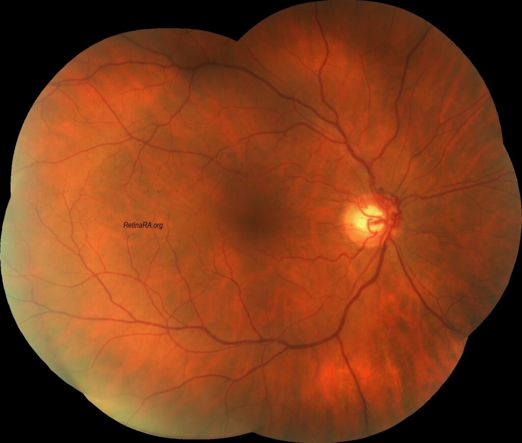
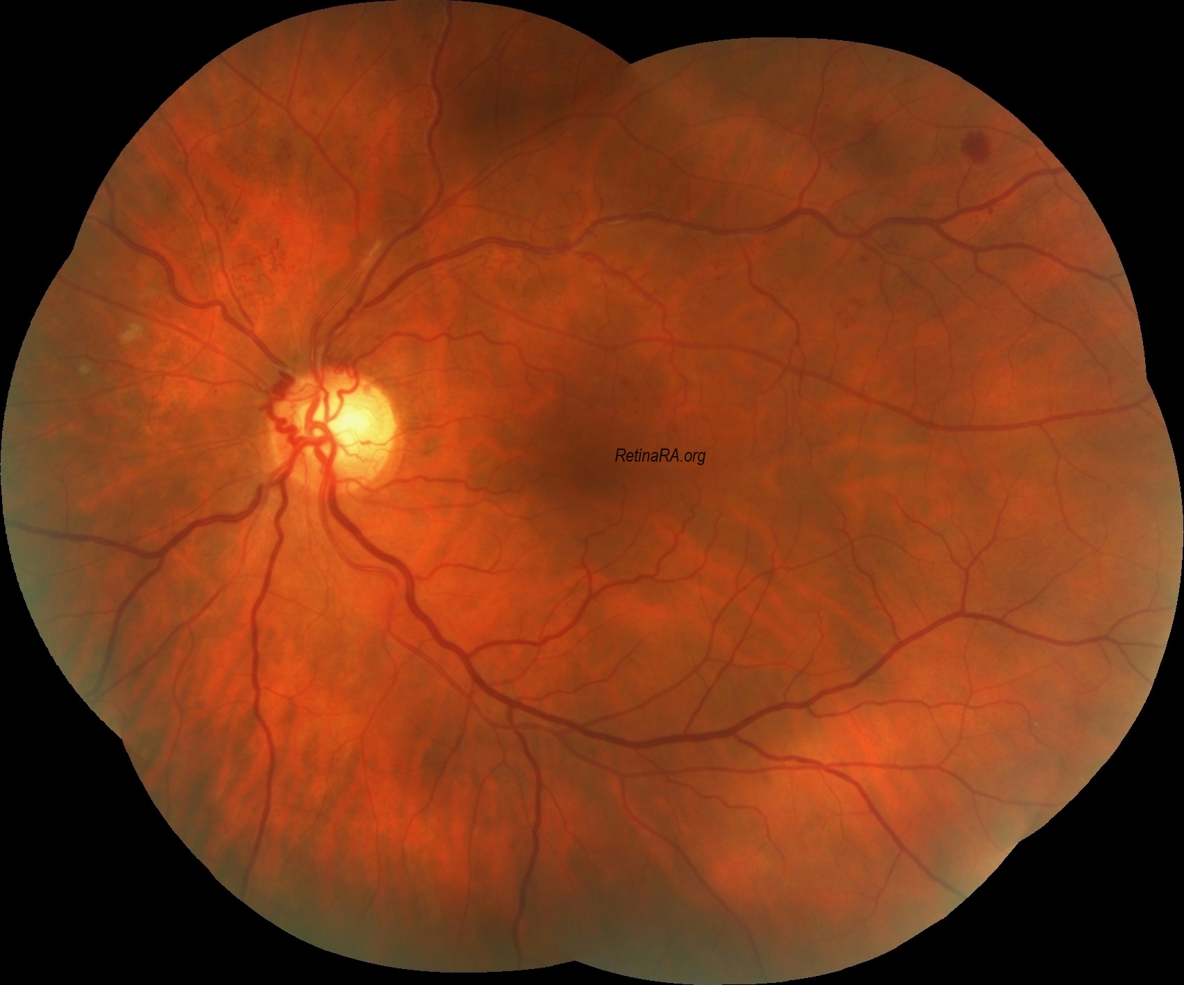
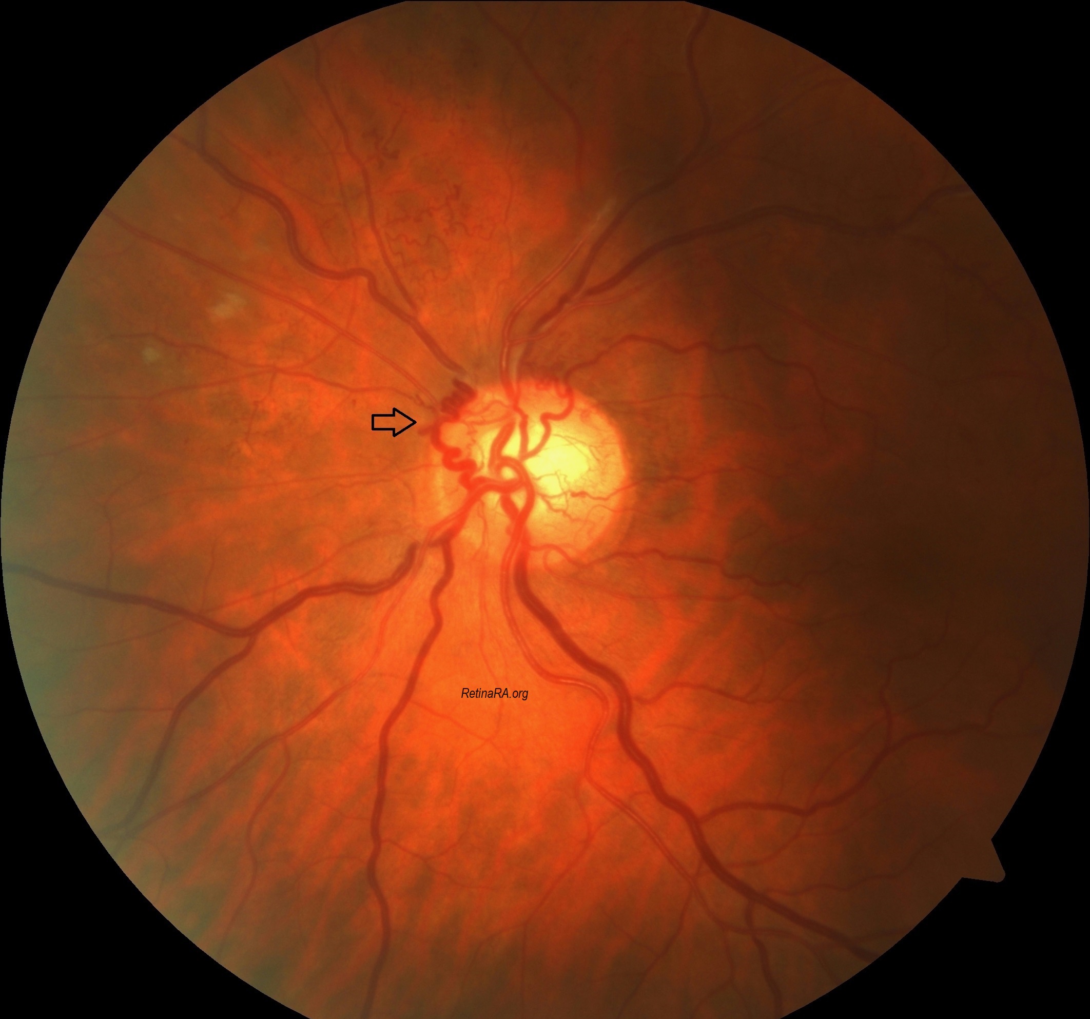
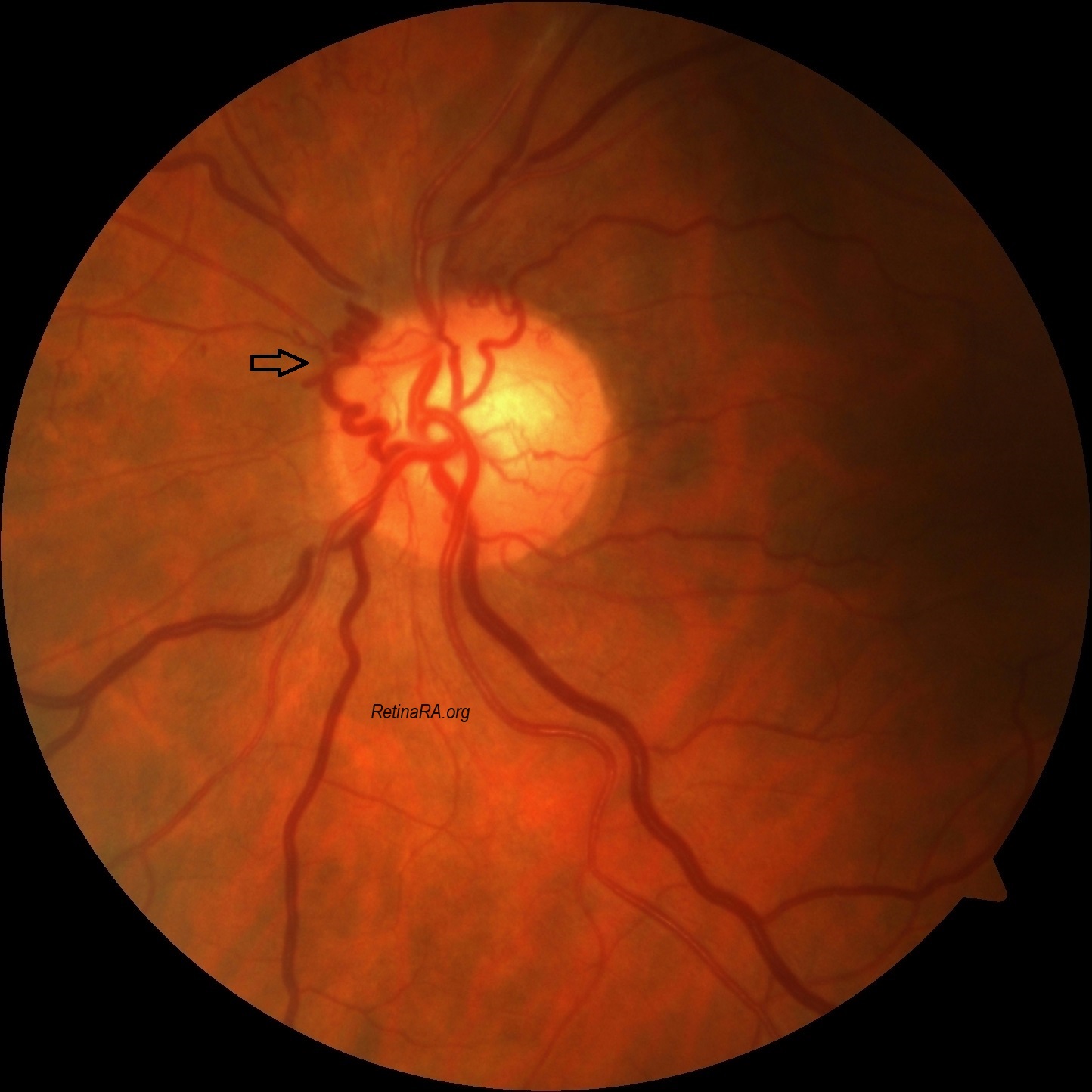
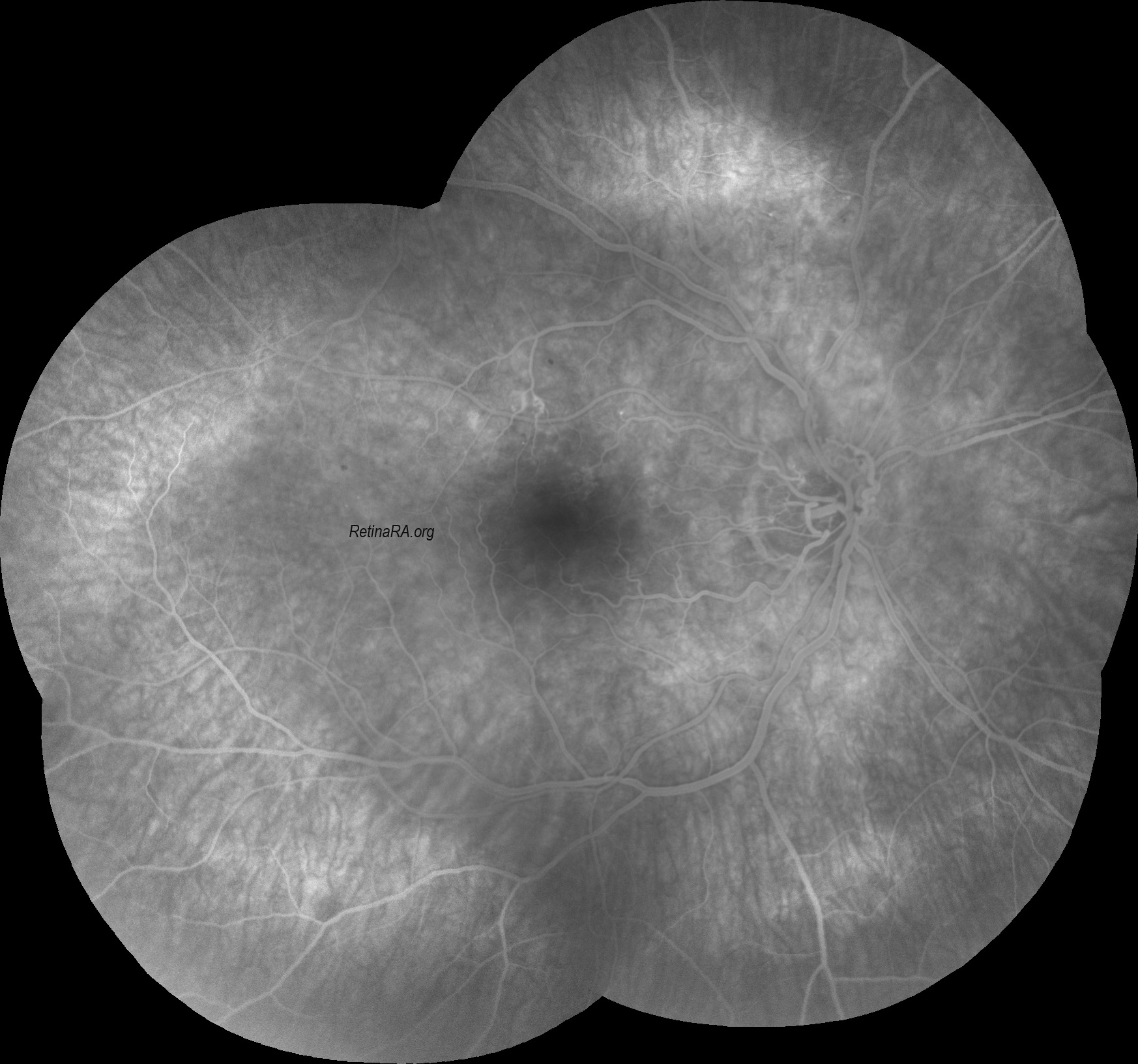
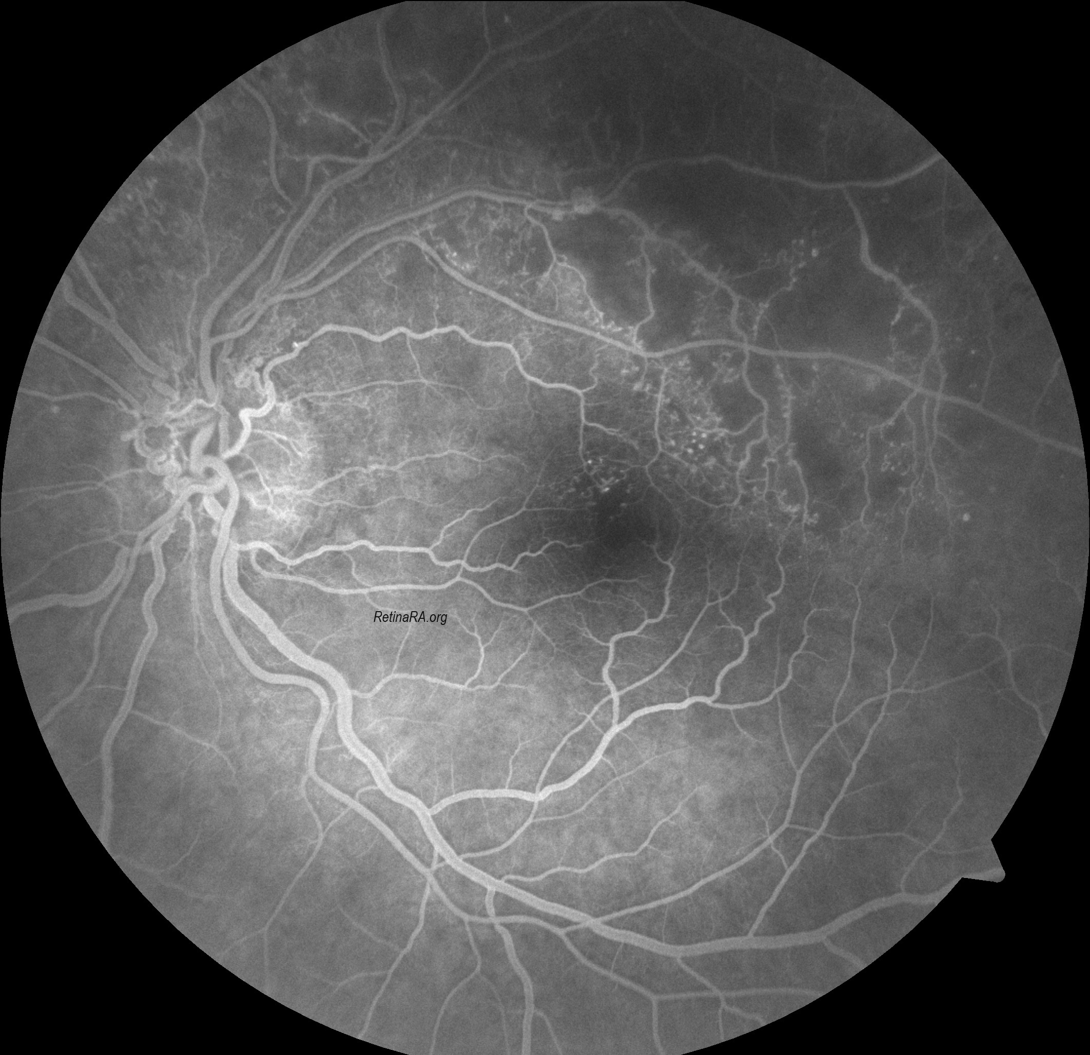
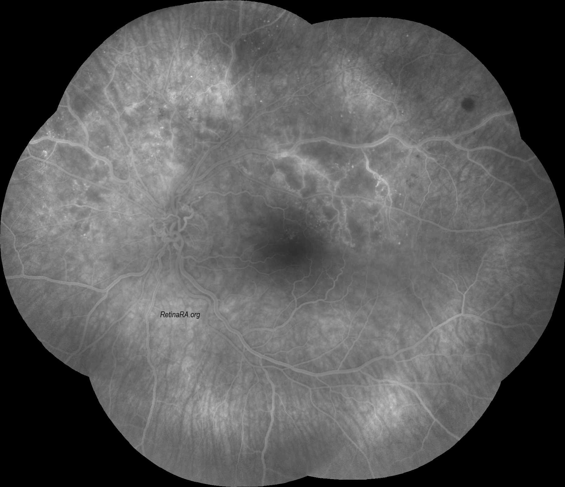
Optociliary shunt vessels, also known as retinochoroidal shunt vessels of the optic disc or retinochoroidal collaterals, are collateral vessels on the optic nerve, connecting the choroidal and retinal circulations. They are associated with a number of conditions including central retinal vein occlusion (CRVO), optic nerve sheath meningioma, chronic glaucoma and chronic papilledema.
Optociliary shunt vessels can be classified as congenital or acquired. Congenital cases are rare. They represent a vascular malformation connecting the retinal and choroidal venous circulations. Acquired optociliary shunt vessels usually occur in association with ophthalmic conditions causing impaired retinal venous outflow.
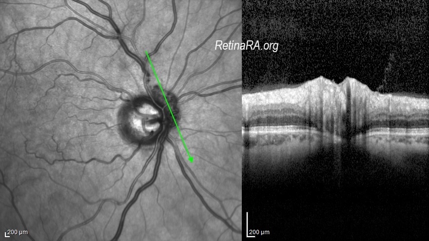
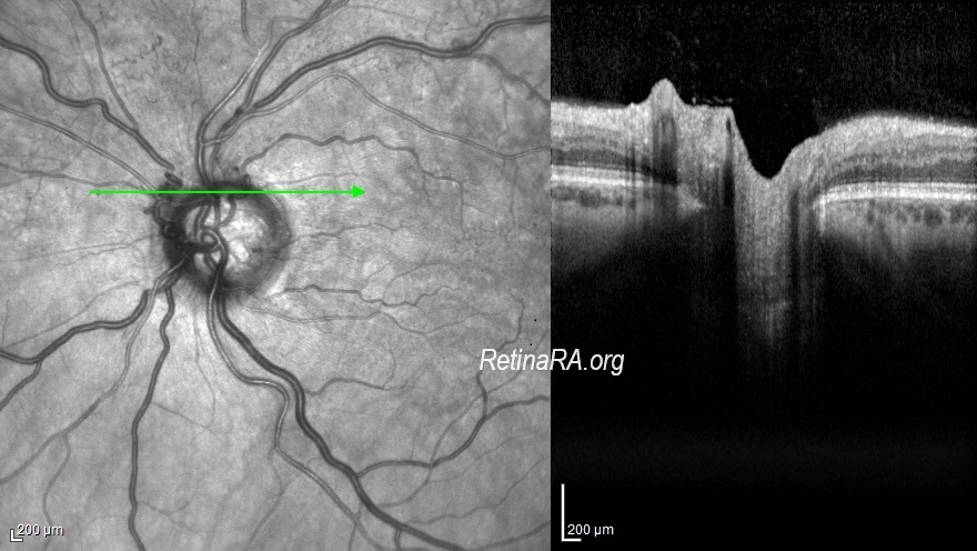
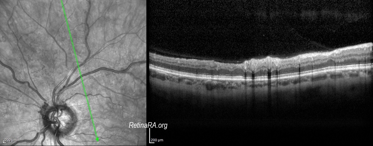
Optociliary shunt vessels are diagnosed on ophthalmic fundus examination, appearing as tortuous vascular loops that begin and end on the disc. Flow within these shunts can be documented in indocyanine green angiography, during the venous phase of fluorescein angiography, or with optical coherence tomography angiography. They must be distinguished from disc neovascularization since neovascularization requires prompt laser photocoagulation whereas shunt vessels can protect against retinal ischemia. Optociliary shunt vessels are generally appear loopy and thick and do not cause leakage on fluorescein angiography.
The clinical approach must include a complete ophthalmic history, especially regarding prievious CRVO, because this the most common association. Fundus examination may reveal some obvious etiologies, such as signs of CRVO, optic disc drusen, or a cupped optic nerve suggestive of glaucoma. Otherwise, ophthalmologists must investigate the causes of chronic compression of the optic nerve, such as optic nerve sheath meningioma or conditions with intracranial hypertension. In patients with papilledema, optociliary vessels might help discern the chronicity of the condition, as they emerge only in the setting of chronic papilledema.
Credit: Kemal Tekin, M.D., from Ulucanlar Eye Training and Research Hospital
Instagram accounts: @retina.academy and @dr.kemaltekin