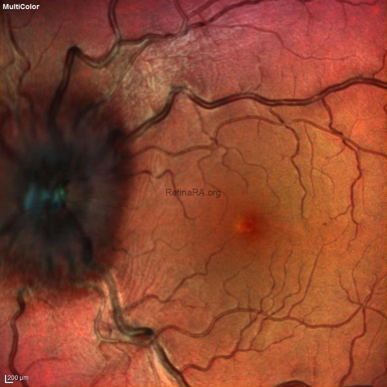
The patient presented with severe vision loss in the left eye. At the time of presentation, visual acuity was 2 meters counting fingers. Optic disc edema appears gray-white at the optic disc borders in color fundus photography. In multicolor imaging, edema is observed as black. Fundus autofluorescence imaging also shows hypoautofluorescence in areas with retinal nerve fiber edema. Late-stage fluorescein angiography reveals leakage in the optic disc. The patient was given 3 days of IV pulse and subsequently oral steroid treatment which was slowly tapered. On the 15th day of treatment, optic disc edema decreased and visual acuity increased to 20/30.
Credit: M. Giray Ersoz, MD, FEBO
Biruni University School of Medicine, Department of Ophthalmology, Istanbul, Turkey
Instagram accounts: @retina.review and @retina.dr.girayersoz
Color fundus photography of optic neuritis
Multicolor imaging of optic neuritis

Fundus autofluorescence imaging of optic neuritis

Early and late phase fluorescein angiography of optic neuritis


Color fundus photograph of optic neuritis on the 15th day of treatment 