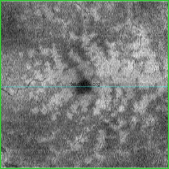
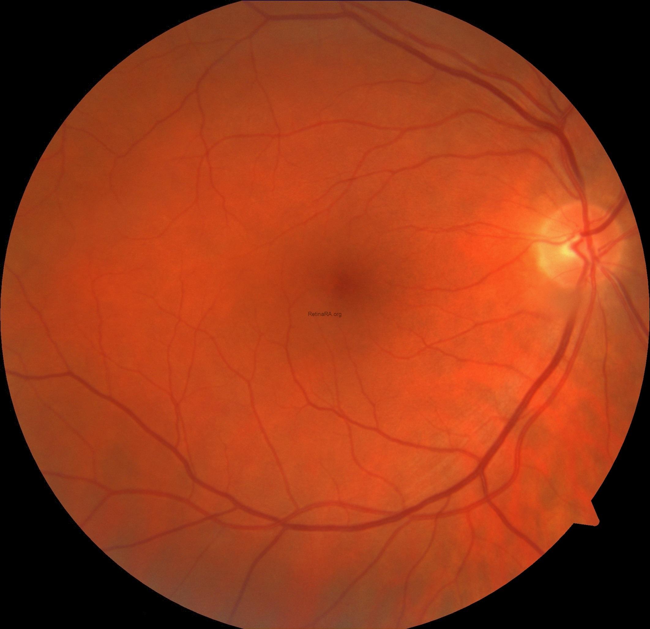
A 72-year-old male patient with Paracentral acute middle maculopathy (PAMM) and Prominent middle limiting membrane (p-MLM) sign secondary to non-ischemic (partial, incipient, Venous stasis retinopathy, etc) central retinal vein occlusion (CRVO). His visual acuity was 20/20 in the right eye and 20/50 in the left eye. Vascular tortuosity and engorgement, superficial retinal hemorrhage, and slight perivenuler whitening around fovea are seen on fundus photography (notice the difference between the two eyes). OCT B-scan shows hyperreflective band-like PAMM lesions and p-MLM sign. En-face OCT demonstrates perivenuler fern-like pattern of PAMM lesions.
PAMM is an OCT finding consisting of characteristic hyperreflective band-like lesions involving the inner nuclear layer that can complicate up to 5.2% of non-ischemic CRVOs. These lesions develop in response to ischemia of the deep retinal capillary plexuses. Prominent middle limiting membrane (p-MLM) sign,” a hyperreflective line in the inner synaptic portion of the outer plexiform layer on OCT B-scan images, indicates acute ischemic retinal damage. The p-MLM sign could be seen on OCT in 94% of ischemic and 66% of nonischemic RVO.
Credit: M. Giray Ersoz, MD, FEBO
Biruni University School of Medicine, Department of Ophthalmology, Istanbul, Turkey
Instagram accounts: @retina.review and @retina.dr.girayersoz
Color Photography of Non-ischemic CRVO + PAMM
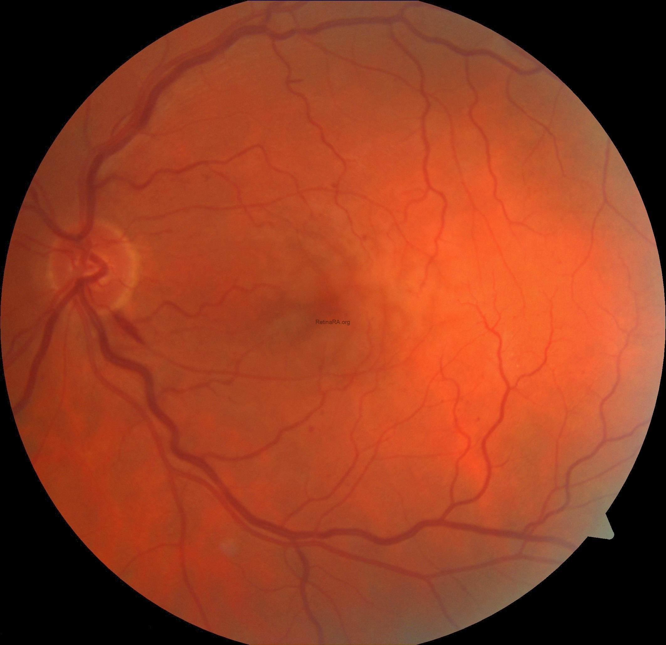
Color Photography of The Fellow Eye (Right Eye)
 Red-free Photography of Non-ischemic CRVO + PAMM

OCT B-Scan of Non-ischemic CRVO + PAMM + P-MLM Sign

En-face OCT of Non-ischemic CRVO + PAMM
