
A 40-year-old male presented to the outpatient clinic for a routine eye examination. Best-corrected visual acuity (BCVA) was 20/20 in both eyes, and intraocular pressures were within normal limits. Anterior segment examination was also unremarkable.
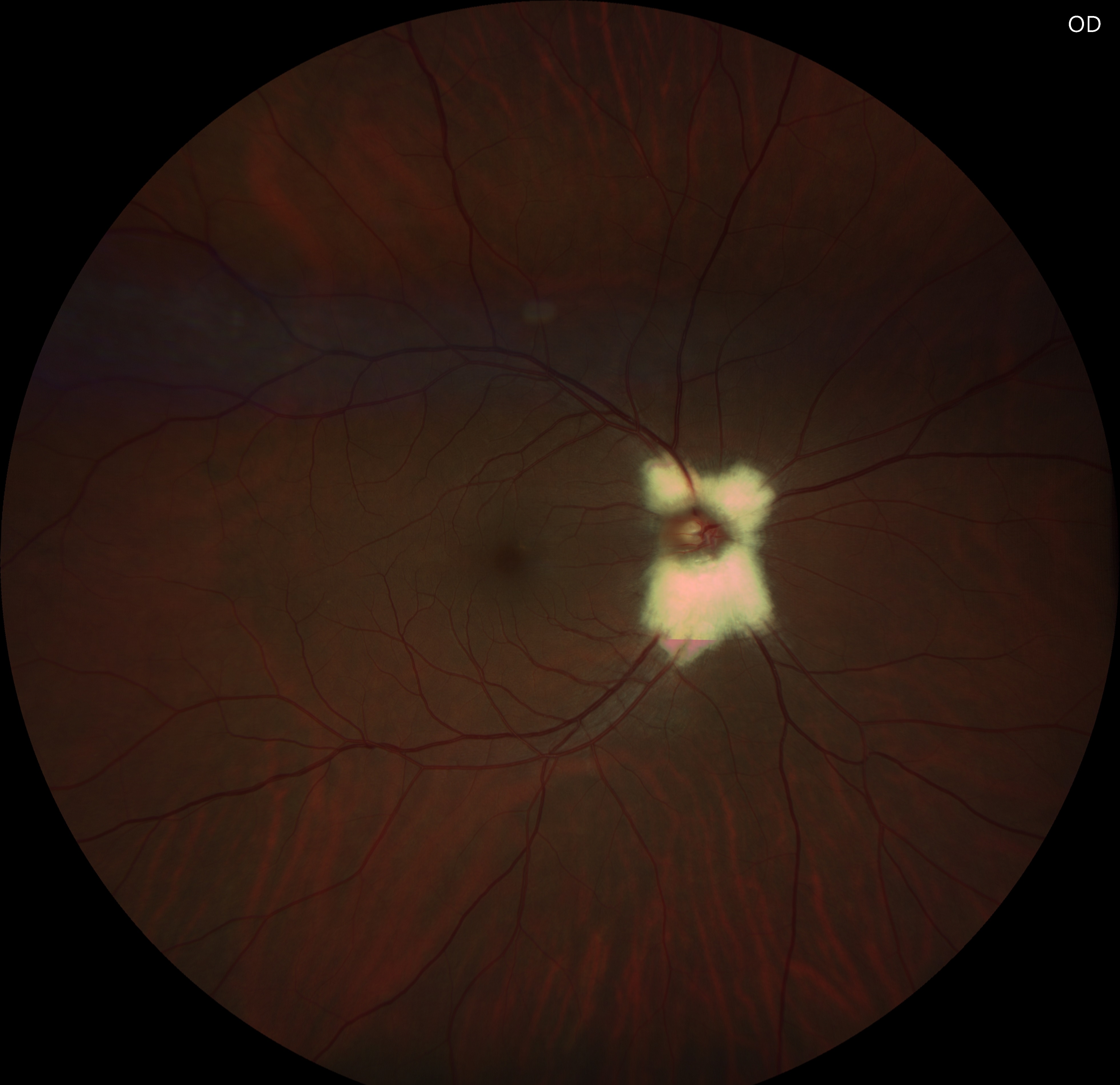
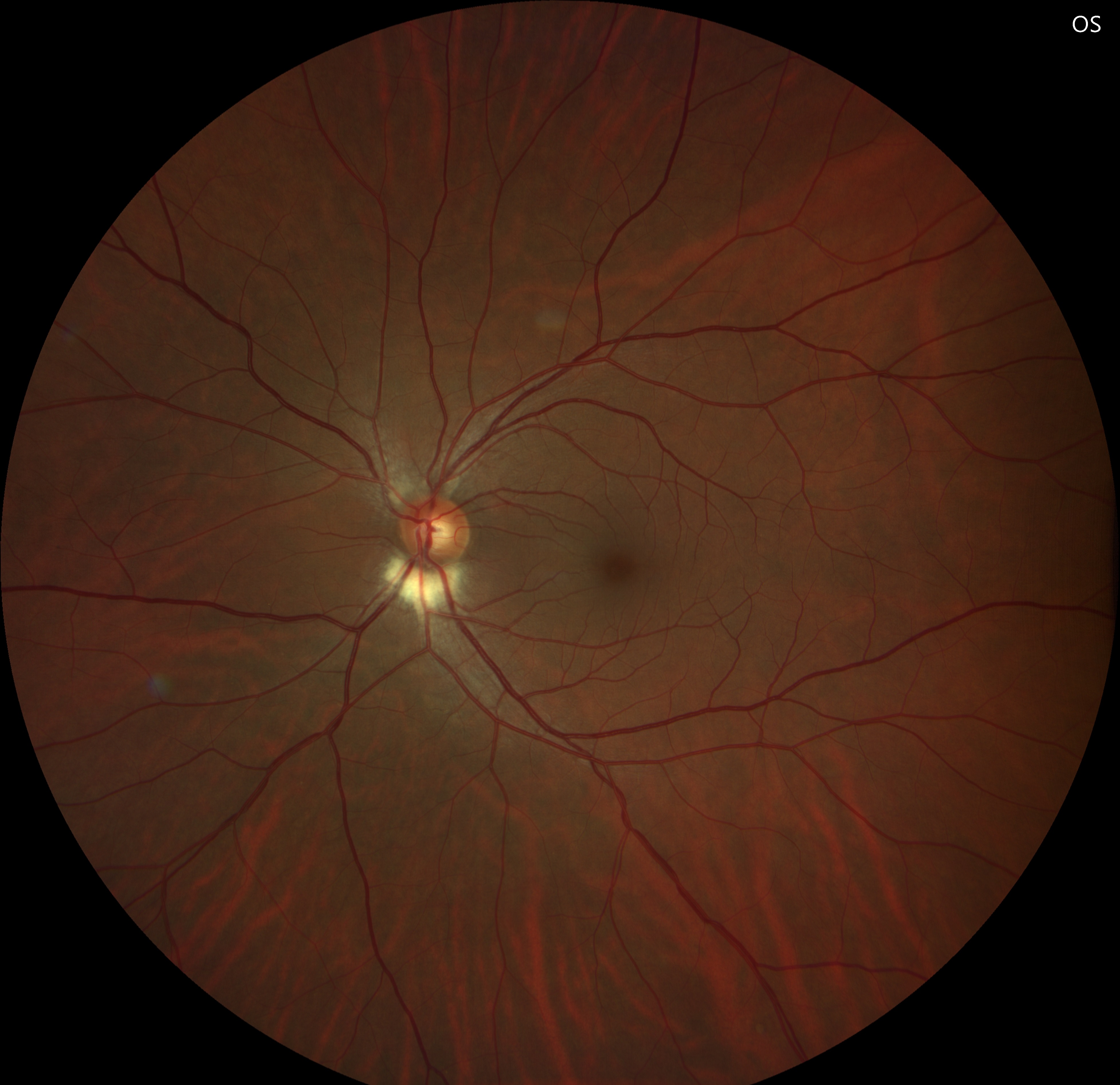
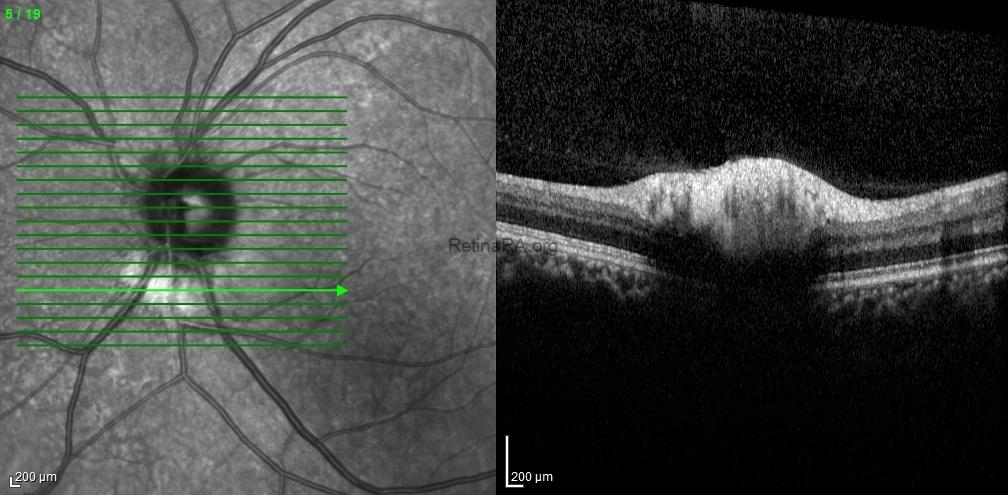
Fundus examination revealed a peripapillary, white-gray, striated patch with feathered borders, approximately one disc diameter in the right eye and half a disc diameter in the left eye.
Optical coherence tomography scans through these lesions demonstrated a thickened and hyperreflective retinal nerve fiber layer.
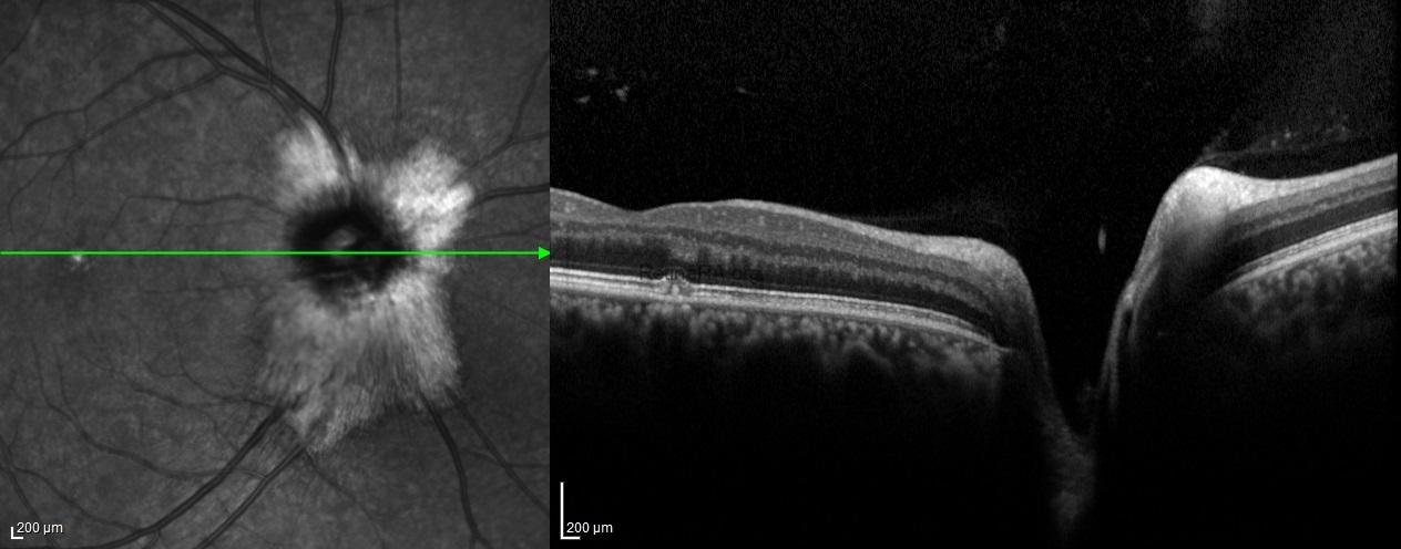
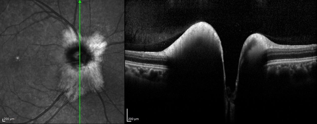
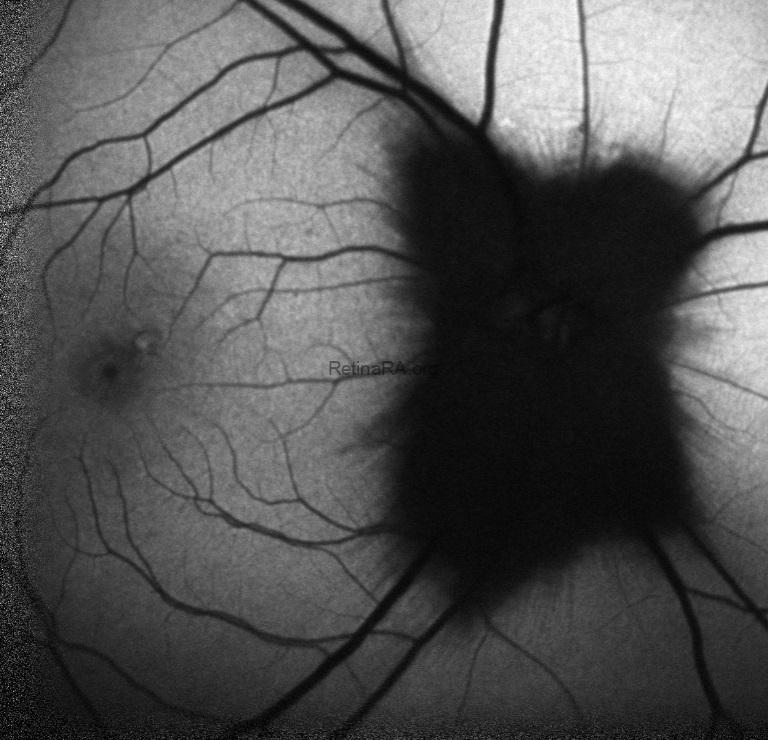
Fundus autofluorescence showed corresponding hypoautofluorescence at the lesion sites.
Myelinated retinal nerve fibers are a rare, benign finding in the eye. Normally, retinal nerve fibers are unmyelinated, but in MRNF, myelin extends into the retina, appearing as white or gray striated patches near the optic disc. They are usually discovered incidentally during routine eye exams and are typically asymptomatic. While vision is often normal, MRNF can sometimes be associated with high myopia or amblyopia. Optical coherence tomography (OCT) shows a thickened, hyperreflective retinal nerve fiber layer corresponding to the lesion. No treatment is needed, but regular monitoring is recommended to rule out other retinal pathologies.
Credit: Kemal Tekin, M.D., from Ulucanlar Eye Training and Research Hospital
Instagram accounts: @retina.academy and @dr.kemaltekin