During embriologic life, there are 2 trunks of the central retinal vein lying on either side of the central retinal artery; one of them usually disappears before birth, leaving the central retinal vein as a single trunk. However, in 20% of eyes, both trunks persist during postnatal life as a congenital abnormality. Occlusion of one of these two trunks in the optic nerve causes the development of hemi-retinal vein occlusion or hemi-central retinal vein occlusion (CRVO).
When branch retinal vein occlusion (BRVO) involves half of the retina (also called hemispheric retinal vein occlusion), it is sometimes confused with hemi-CRVO, and involvement of one quadrant of the retina by hemi-CRVO has been misdiagnosed as BRVO. Some ophthalmologists tend to consider hemispheric retinal vein occlusion and hemi-CRVO as the same disease or use the terms interchangeably. However, the two are very different clinical situations.
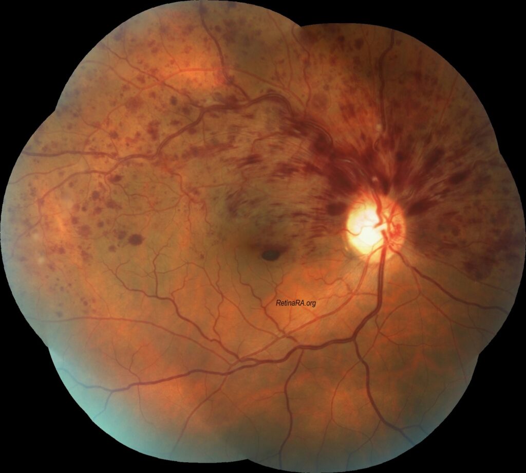
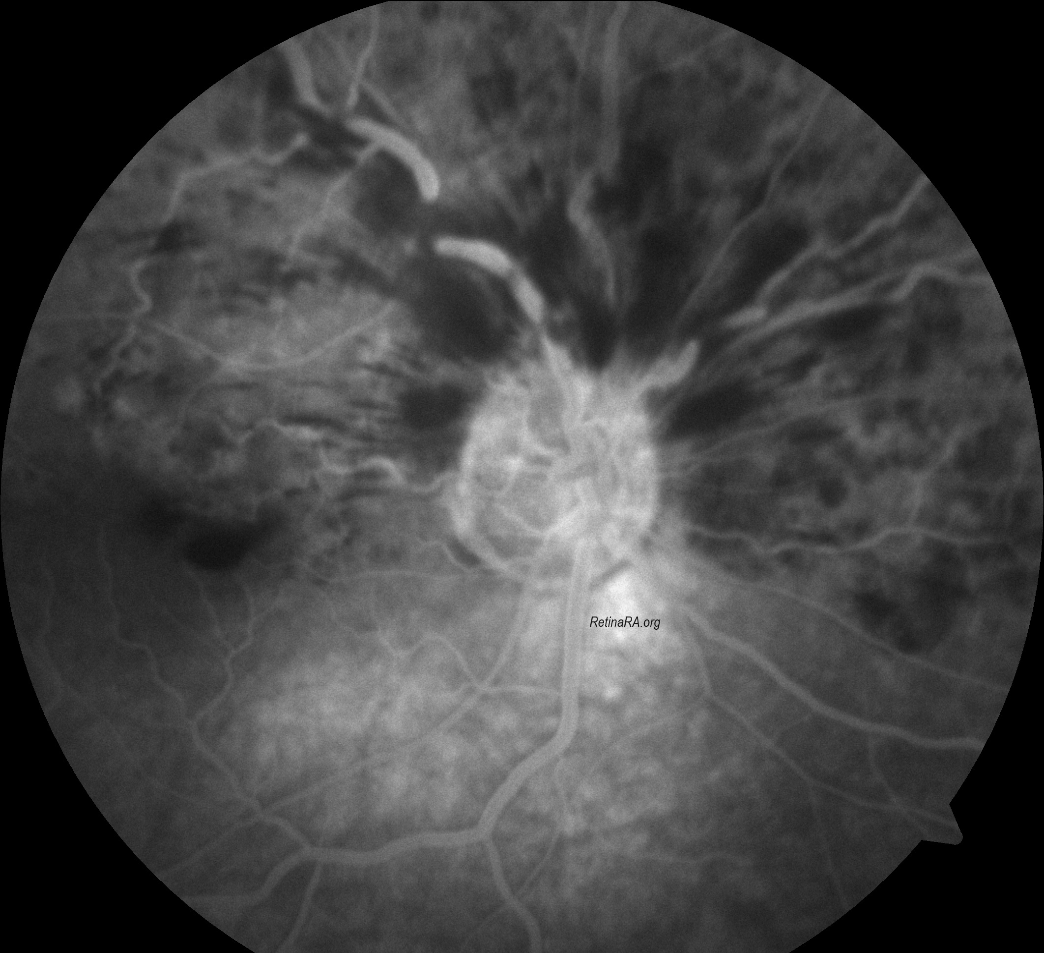
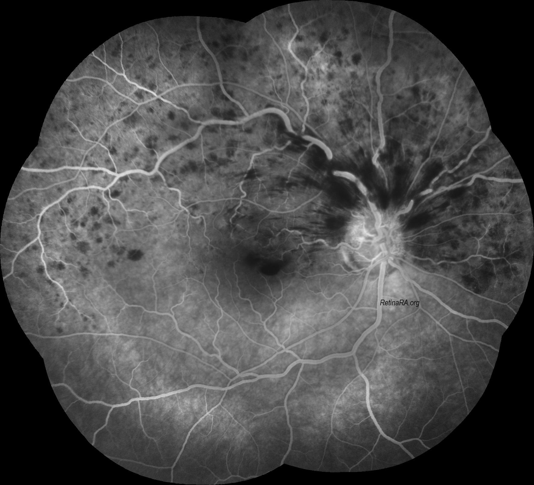
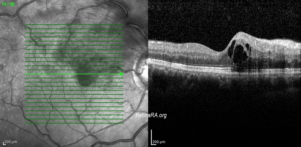
Hemi-CRVO has been shown to be a distinct entity that is clinically and pathogenetically closely related to CRVO and unrelated to BRVO due to fundamental differences between the two. Hemi-CRVO clinically presents as either venous stasis retinopathy or as hemorrhagic retinopathy, usually involving one half of the retina, although occasionally it may involve one third to two thirds of the retina. The clinical features of venous stasis retinopathy and hemorrhagic retinopathy caused by hemi-CRVO are identical to those caused by CRVO. Visual prognosis seems to be better in hemi-CRVO than in total CRVO.
Credit: Kemal Tekin, M.D., from Ulucanlar Eye Training and Research Hospital
Instagram accounts: @retina.academy and @dr.kemaltekin
