
A 60-year-old man with a silent medical history comes for a second consultation after a misdiagnosis of central serous chorioretinopathy.
Cuticular drusen, also known as basal laminar drusen, were first described by Gass (1977). They are small, round, yellowish lesions measuring approximately 50–75 μm in diameter. These lesions are typically bilateral and tend to increase in number over time, often clustering and coalescing to form pigment epithelial detachments (PEDs). Cuticular drusen are usually observed in younger adults (40–60 years) and have a strong genetic predisposition, particularly associated with variants in the complement factor H (CFH) gene (Duvvari et al., 2015).
Imaging Characteristics
* Optical Coherence Tomography (OCT):
Displays triangular or dome-shaped elevations of the RPE–basal lamina complex, with their bases on Bruch’s membrane and apices directed toward the retina, producing a characteristic “saw-tooth” configuration (Fragiotta & Scuderi, 2021).
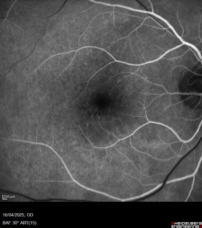
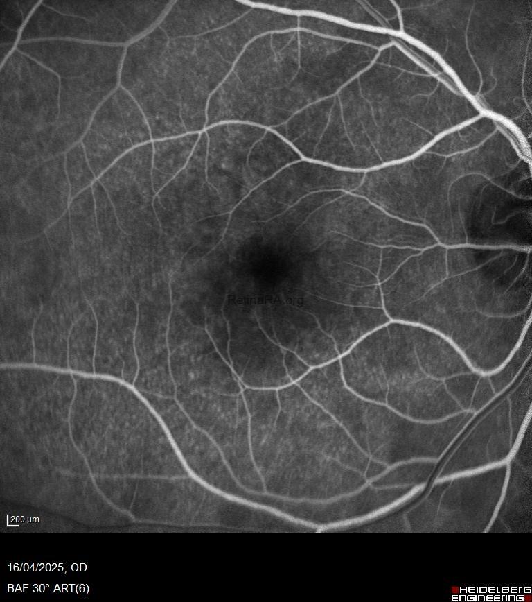
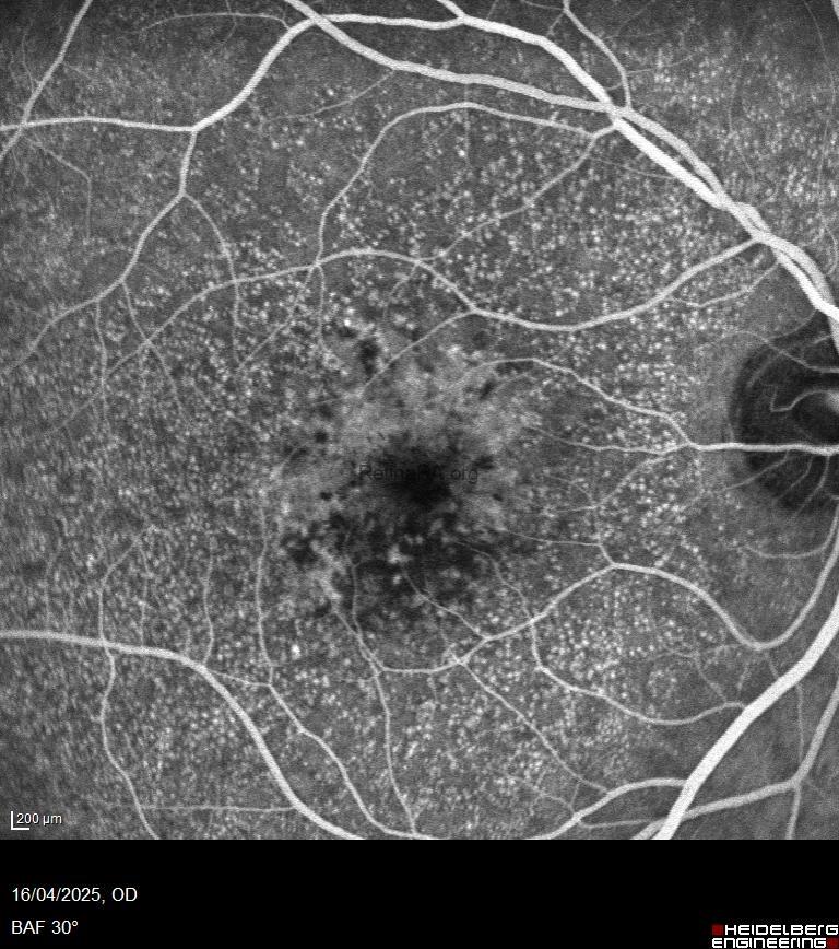
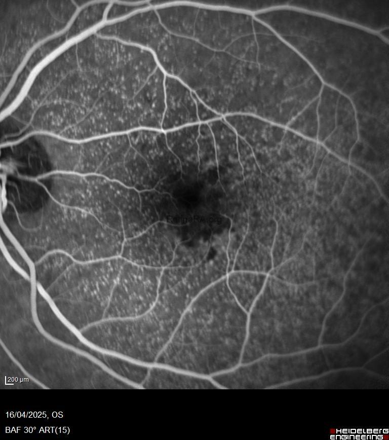
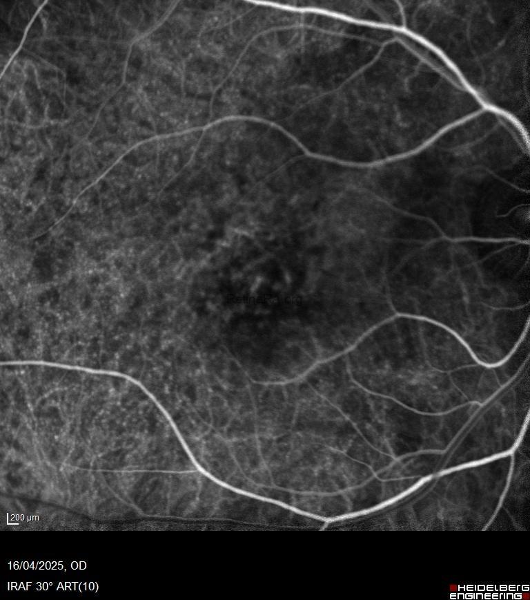
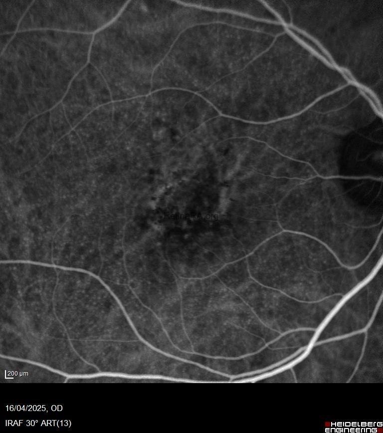
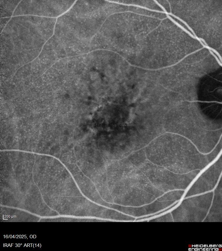
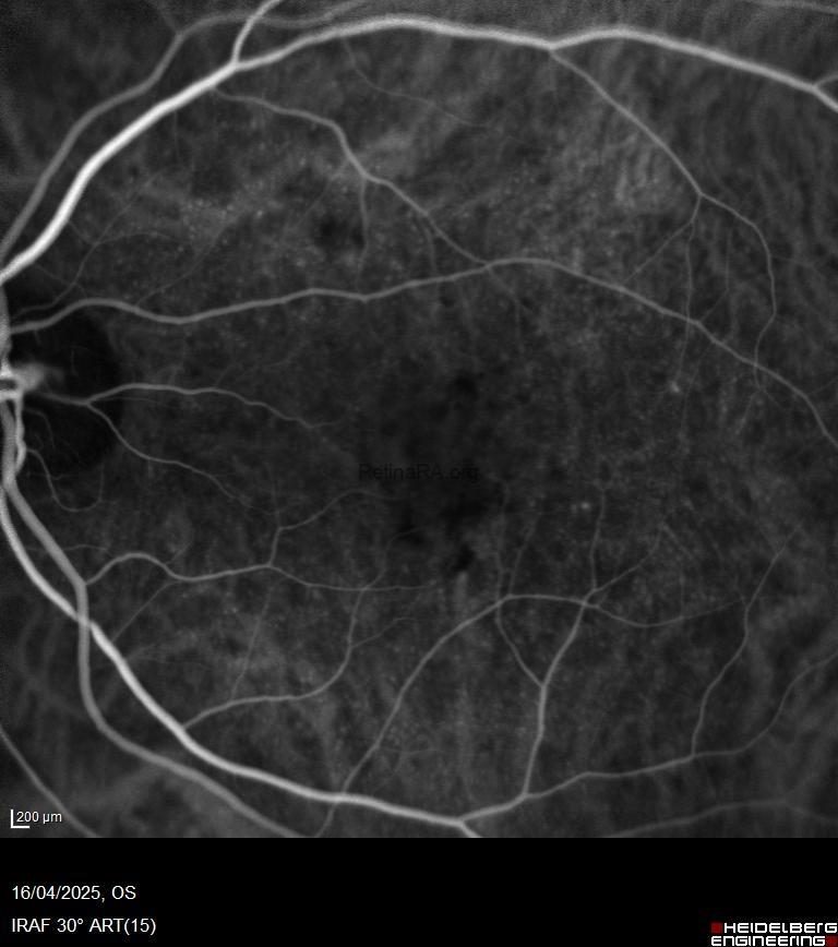
* Fundus Fluorescein Angiography (FFA/FAG):
Exhibits a characteristic “starry-sky” or “milky-way” pattern, with numerous, small, round hyperfluorescent lesions appearing during the intermediate or arteriovenous phase.
* Indocyanine Green Angiography (ICGA):
Shows early hypercyanescence surrounded by a hypocyanescent halo, which becomes uniformly hypercyanescent in the late phases.
* Fundus Autofluorescence (FAF):
Lesions appear hypoautofluorescent, often bordered by a ring of hyperautofluorescence.
Clinical Associations and Complications
The most notable clinical association is the presence of acquired vitelliform lesions (AVLs) in the foveal region (Gass, 1985). Eyes with cuticular drusen are also at increased risk for developing large drusen and choroidal neovascularization (CNV), particularly type 1 CNV.
* On FFA and ICGA, vitelliform material shows hypofluorescence ed hypocyanescent due to blockage.
* On OCT, it appears as hyperreflective subretinal material with underlying hyporeflective fluid.
In the absence of other age-related macular degeneration (AMD) features, such presentations suggest adult-onset foveomacular dystrophy (AOFD).
With disease progression, AVLs often resorb, and drusen may disappear, but this typically results in retinal pigment epithelium (RPE) atrophy.
Key References
* Gass JD. Arch Ophthalmol. 1977;95:335–339.
* Gass JD. Arch Ophthalmol. 1985;103:615–623.
* Duvvari MN et al. Invest Ophthalmol Vis Sci. 2015;56:324–330.
* Fragiotta S, Scuderi G. Surv Ophthalmol. 2021;66(4):596–610.
Author
Giovanni Di Fiore, M.D. Naples @giodifiore
Claudio Iovino, M.D. Naples @claudioiovino