A 14-year-old boy was referred for the evaluation of decreased vision in both eyes. He has been followed with diagnosis of X-linked retinoschisis in another clinic and using topical dorzolamide. The boy was a product of consanguinity marriage in which the parents of the patient were a second degree relative. On ocular examination, BCVAs were 20/30 in both eyes and IOPs were within normal limits. Anterior segment examinations of both eyes were also unremarkable.
Dilated fundus examination showed bilateral maculopathy with multifocal subretinal white-yellow deposits in the macula and posterior pole of both eyes.
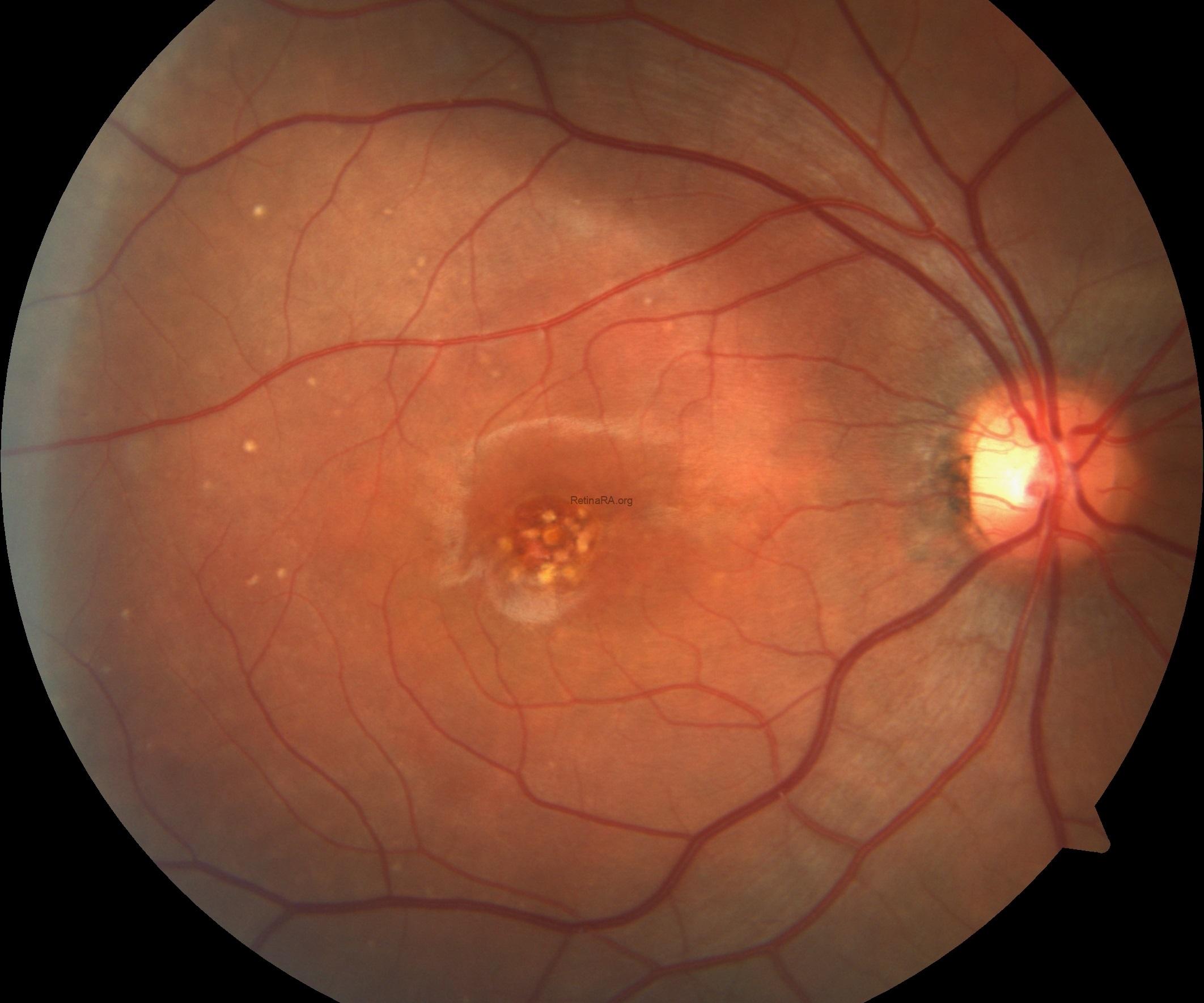
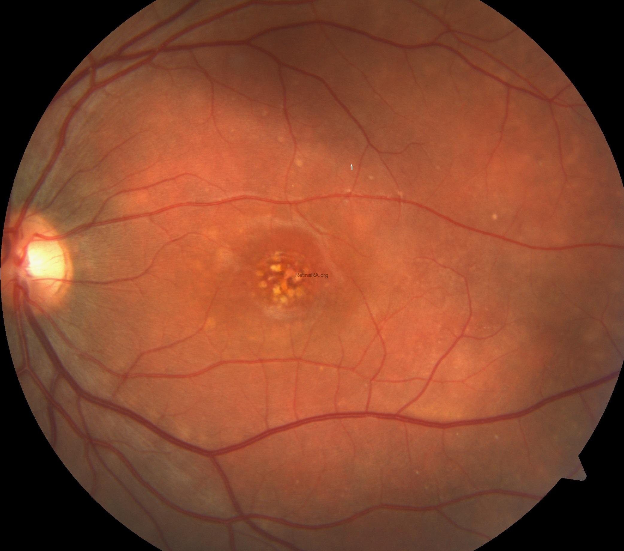
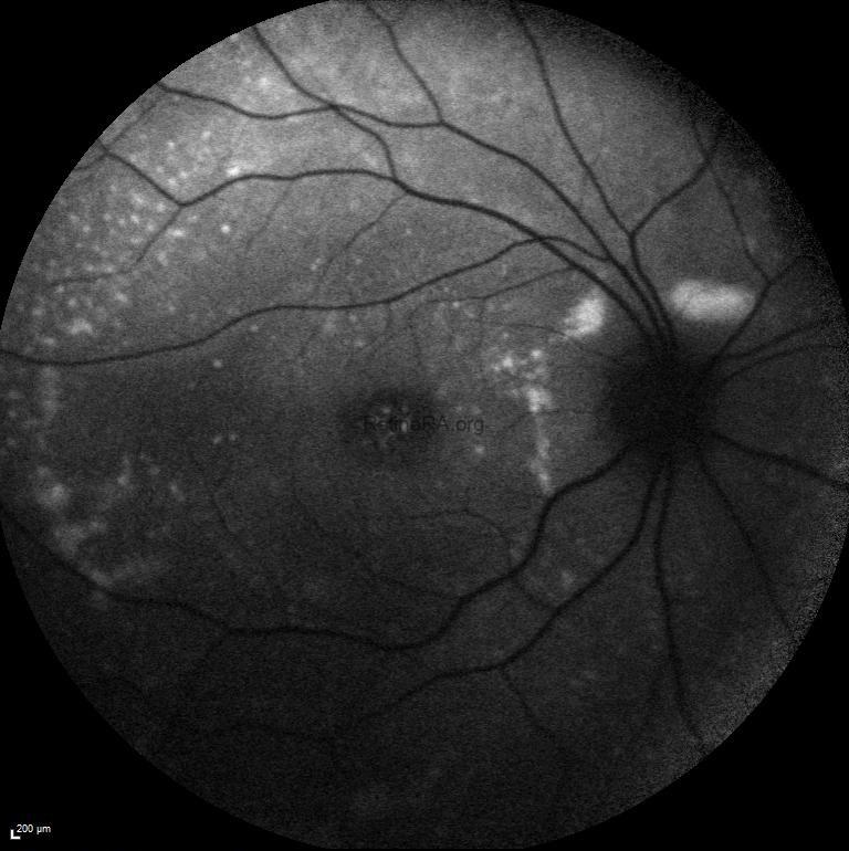
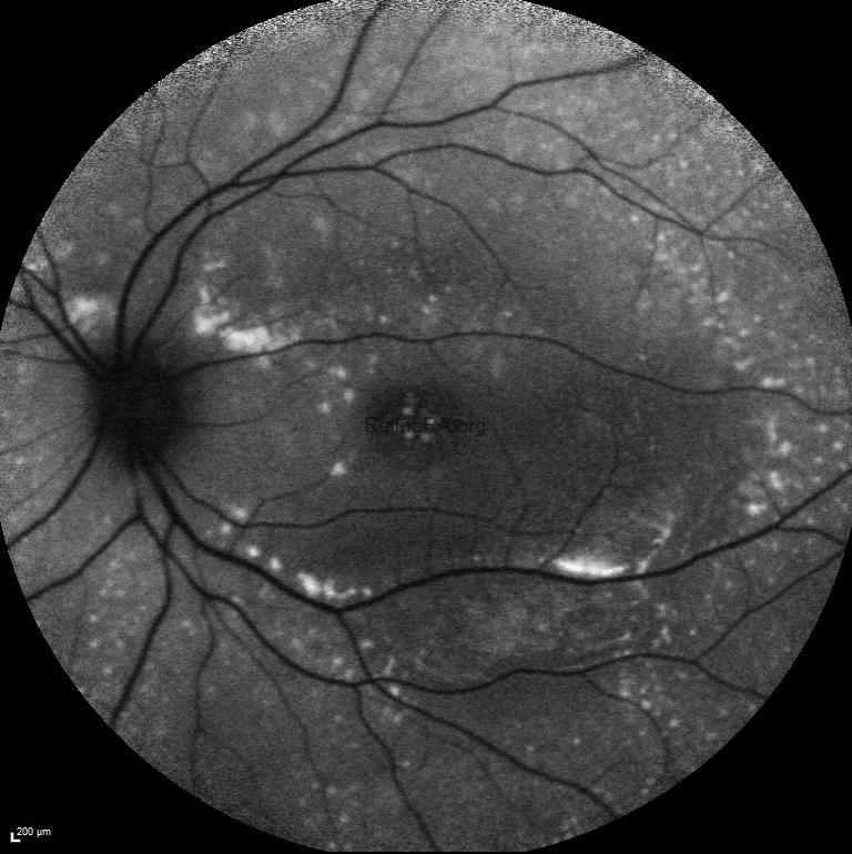
Fundus autofluoresce imaging demonstrated widespread hyperfluorescent foci corresponding to the subretinal deposits in both eyes.
Vertical and horizontal optical coherence tomography scans passing through the fovea revealed serous retinal detachment, retinoschisis with intraretinal cysts, brush border appearance caused by elongation of the outer segments of photoreceptors, and hyperreflective dome-shaped deposits at the level of retinal pigment epithelium in both eyes.
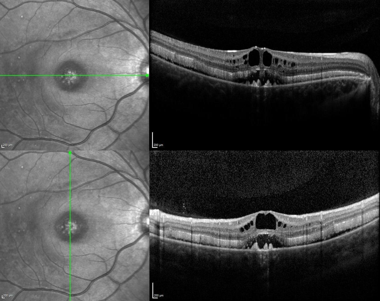
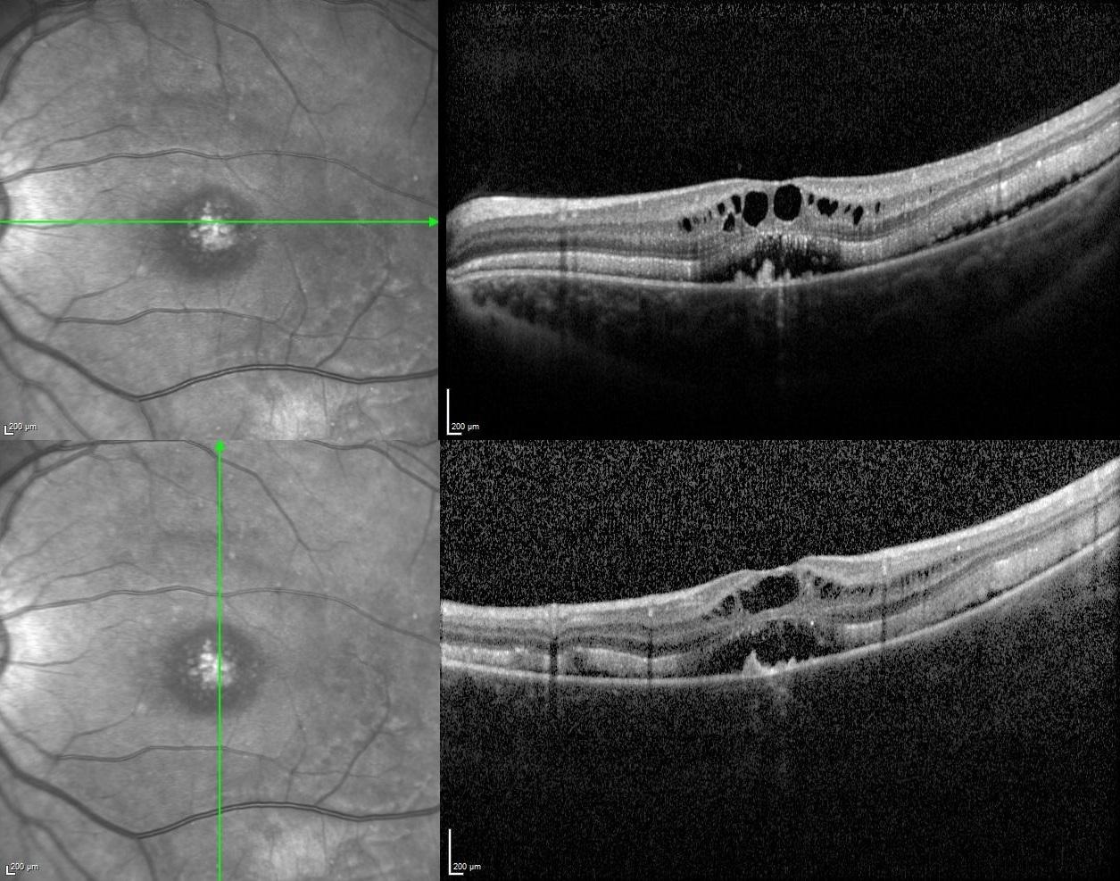
According to the multimodal imaging characteristics, the preliminary diagnosis was ARB and electrooculogram (EOG) was performed to confirm the diagnosis. EOG showed the absence of light rise in which Arden ratios were detected as 1.46 in the right and 1.51 in the left eye. For the definitive diagnosis, genetic analysis was performed and compound heterozygous mutation in BEST1 gene was detected.
Autosomal recessive bestrophinopathy was first introduced in 2008 and its clinical manifestations include decreased visual acuity because of subretinal fluid or macular edema, characteristic retinopathy with the punctate flecks and vitelliform deposits, an absent or severely reduced EOG, in addition to hyperopia and a shallow anterior chamber increasing the risk of angle-closure glaucoma. Since ARB can be presented with a wide spectrum of ocular abnormalities that may not be easily diagnosed, some misdiagnosis can be made.
Credit: Kemal Tekin, M.D., from Ulucanlar Eye Training and Research Hospital
Instagram accounts: @retina.academy and @dr.kemaltekin
