A 47-year-old male patient presented with decreased vision in his left eye. He also had characteristic skin lesions involving the neck and axilla. Best-corrected visual acuity was 1.0 in the right eye and 0.1 in the left eye. Intraocular pressures were within normal limits, and anterior segment examination was unremarkable in both eyes.
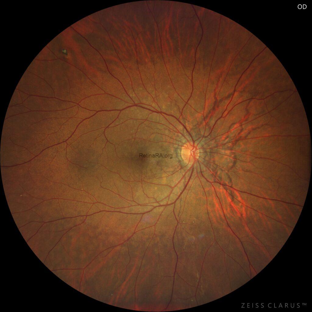
Fundus photography revealed bilateral angioid streaks radiating from the optic disc, appearing as irregular, reddish-brown linear lesions. Additional findings included a peau d’orange appearance of the fundus and scattered comet-like peripheral lesions. In the left eye, a choroidal neovascular membrane was detected in the macular region, accompanied by a choroidal rupture–like lesion. Overall, these findings were consistent with angioid streaks complicated by secondary choroidal neovascularization.

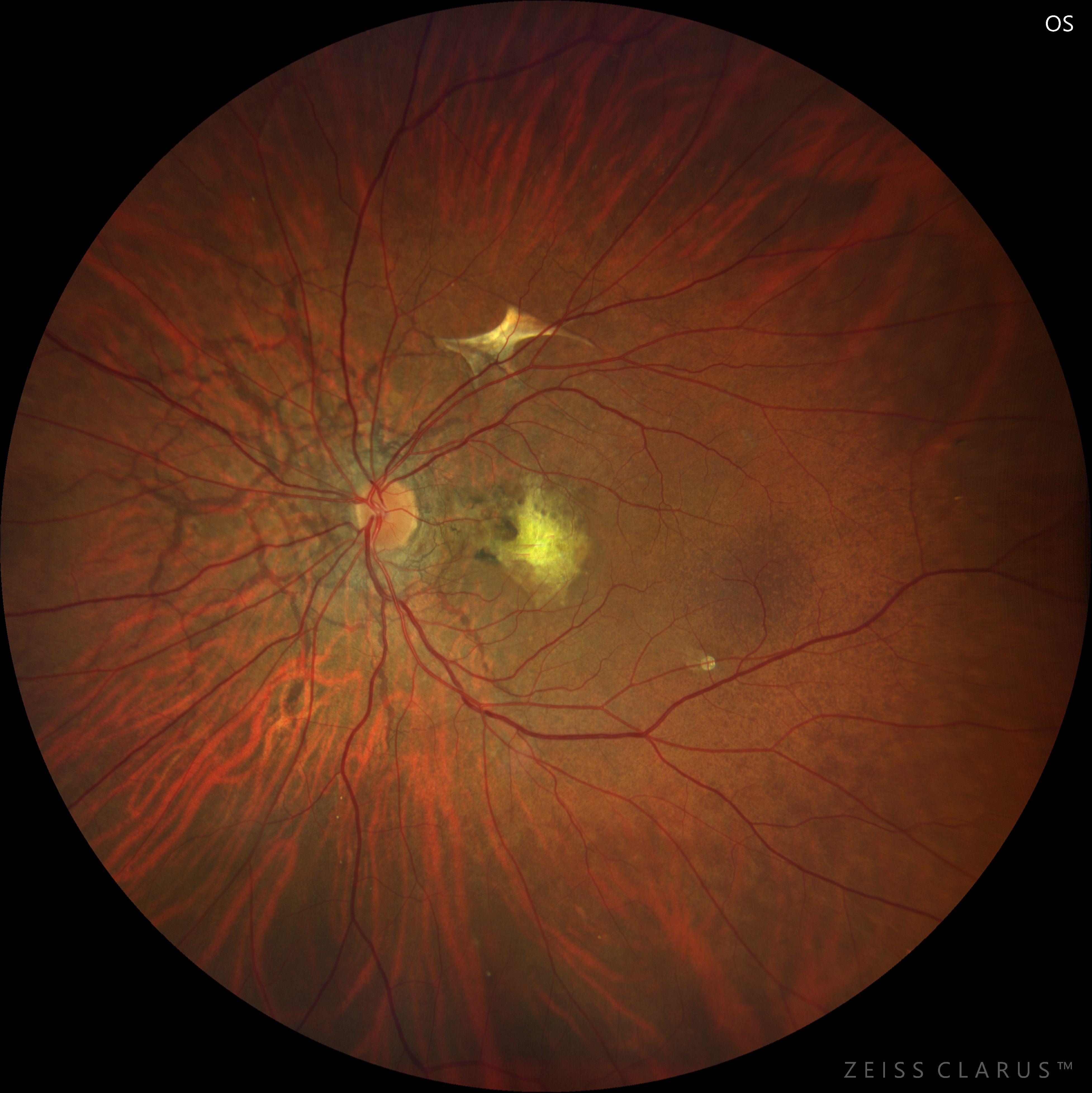
Angioid streaks are crack-like dehiscences of a pathologically calcified Bruch’s membrane, typically radiating from the optic disc. They are often bilateral and may be associated with systemic conditions such as pseudoxanthoma elasticum, Paget disease, and hemoglobinopathies. Funduscopic findings include reddish-brown linear streaks, peau d’orange appearance, and peripheral comet lesions. The most vision-threatening complication is choroidal neovascularization (CNV), which commonly affects the macula and leads to sudden visual loss. Early recognition, patient counseling to avoid ocular trauma, and prompt detection of CNV are crucial, as timely anti-VEGF therapy can preserve vision.
Credit: Kemal Tekin, M.D., from Ulucanlar Eye Training and Research Hospital
Instagram accounts: @retina.academy and @dr.kemaltekin